

Introduction
High levels of radiation from radioactive sources are harmful to living organisms and can cause irreversible damage, cancer, and even death. Radiation has a permanent or temporary effect on fertility, depending on the dose. Furthermore, the production of sex steroids can be disrupted. In adults, this leads to sex steroid deficiency, and in childhood, the sexual cycle can be disrupted [1].
The testicles are a radiation-sensitive organ due to the high rate of spermatogonia proliferation. Exposure to radiation can cause reversible or irreversible damage to the reproductive system [2]. Radiation can cause various defects in spermatogenesis, leading to reduced sperm count and an increased incidence of sperm disorders [3, 4]. Oxidative stress leads to loss of sperm DNA and a decrease in the fertilizing ability of these cells due to bystander damage to protein and fat components in the plasma membrane of sperm [5, 6].
One of the most important strategies for reducing the damage caused by ionizing radiation is the use of radioprotectors. These substances are administered before irradiation to reduce damage and cell death from ionizing radiation. The underlying mechanism of radioprotector protection is their role in inhibiting free radicals. This effect protects cells from reactive oxygen species generated by radiation or chemotherapeutic agents [7]. A standard radioprotector must be able to reduce the adverse effects of ionizing radiation and protect normal tissues. Furthermore, it must have low toxicity, adequate efficacy, desirable stability, high uptake, rapid tissue distribution, and no interactions with other drugs. For similar reasons, the use of materials naturally present in the body has attracted considerable attention in this regard [8]. Radiation-protective compounds reduce the negative impact of ionizing radiation on body tissues through various mechanisms, including scavenging free radicals, transferring hydrogen atoms to damaged molecules, causing hypoxia, and reducing the production of reactive and atomic oxygen species, stimulating the proliferation and differentiation of hematopoietic stem cells, increasing the activity of endogenous antioxidants in the body, as well as binding to DNA and increasing its stability [9].
Selenium is a cofactor or activator of the enzyme glutathione peroxidase, which is one of the most powerful natural antioxidants: it promotes the breakdown of lipid peroxidases and hydrogen peroxide. Glutathione peroxidase is one of the most important antioxidants against damage caused by free radicals and is considered a powerful radiation shield [7, 8]. Adequate selenium levels enhance antioxidant activity and support overall health. Selenium is also involved in testosterone production. In addition, selenium is involved in sperm development, and decreased levels lead to atrophy of the seminiferous tubules, reducing sperm production [10]. Long-term selenium deficiency in the diet of guinea pigs reduces sperm concentration and motility and causes the formation of cytoplasmic droplets in sperm [11]. Studies have shown that selenium improves reproductive performance in organisms such as mice, pigs, and sheep [12]. Selenium has been found to protect tissues from oxidative damage and modulate the immune microenvironment, promoting hormone development and expression [13, 14]. Recently, selenium nanoparticles have attracted widespread attention due to their high availability and less toxic effects compared to organic and inorganic forms of selenium. Selenium nanoparticles/nanocomposites and other biocompatible nanoparticles have also been confirmed to have beneficial effects in the treatment of cancer and other diseases. Selenium nanoparticles exhibit high free radical scavenging properties. Nano-selenium is used to enhance selenium efficacy and reduce toxicity [15, 16]. In general, nanomaterials enhance the dispersibility, stability, and availability of bioactive substances [17, 18].
Similarly, probiotics such as Lactobacillus casei Shirota may have a radioprotective effect. Probiotics are generally live bacteria that are beneficial to the body’s health [19]. Some species are strictly anaerobic and have a fermentative metabolism that produces energy through the fermentation of sugars [20]. These bacteria help balance the body’s microbial flora and have a beneficial effect on the immune system [21]. It is worth noting that previous studies have shown colonization of L. casei in the intestinal tract of mice after oral administration [22]. Most notably, this probiotic exerts a corresponding antioxidant and antiapoptotic effect in mouse testes after X-ray irradiation, enhancing the compound’s radioprotective effect [23].
The objective of this study was to investigate the protective role of nano-selenium and L. casei against the effects of radiation therapy on the reproductive and hematological systems. The radioprotective effect of this combination of substances has not been previously assessed. Therefore, we examined the effects of this formula on hematological, biochemical, and hormonal parameters to determine whether it could preserve physiological functions after irradiation.
Material and Methods
Material
Selenium nanopowder was obtained by chemical reduction and purchased from Pishgaman Company, Iran. Nano-selenium was administered orally at a dose of 0.2 mg/kg daily. L. casei (Pasteur Institute of Iran) was cultured in MRS broth at 37 °C under anaerobic conditions for 24–48 h. Bacterial cells were collected by centrifugation (4,000 ×g, 15 min, 4 °C), washed twice with sterile phosphate-buffered saline (PBS, pH=7.4), and resuspended in PBS to a concentration of 1×10⁹ CFU/mL. Viability was confirmed before each administration by plating serial dilutions on MRS agar and counting colonies after 48 h of anaerobic incubation at 37 °C. Each laboratory animal was administered 100 μL (1×10⁸ CFU) orally daily using a sterile 20-gauge needle.
Experimental design and model validation
This study was designed and reported in accordance with ARRIVE (Animal Research: Reporting of In Vivo Experiments) 2.0 guidelines and the principles outlined in the Guide for the Care and Use of Laboratory Animals (8th edition, National Institutes of Health, USA). A total of 64 male Syrian mice (n=8 in each group) were randomly assigned to 8 experimental groups using a computer-generated randomization sequence. The sample size was determined a priori based on a power analysis (80% power, α=0.05) using effect sizes from pilot experiments [23]. Group assignment and outcome assessment were performed by investigators blinded to treatment conditions. All animals were acclimated for 7 days prior to treatment. Health status, body weight, and behavioral signs of distress were monitored daily. No animals were excluded from the analysis, and any morbidity or mortality would have been recorded, but this did not occur. The study protocol (IR.TUMS.MEDICINE.REC.1400.1104) explicitly included measures to minimize pain and stress in the animals, including anesthesia (ketamine/xylazine) before irradiation and euthanasia by decapitation under deep anesthesia at the study endpoint, in accordance with AVMA guidelines.
The 2 Gy X-ray irradiation model was thoroughly validated at the histological, biochemical, cellular, and molecular levels in our pilot study [23]. The same irradiation parameters and injury criteria were employed in the present study, thereby ensuring reproducibility. In other words, the successful induction of X-ray damage was confirmed histologically, biochemically, and by flow cytometry in our previous study using identical irradiation parameters [23], where the X-ray-only group showed a significant decrease in spermatogenesis, antioxidant enzyme activity, and the percentage of viable cells, along with an increase in apoptosis and malondialdehyde levels.
To evaluate the effects of nano-selenium and probiotics on biochemical, hormonal, and hematological parameters in male Syrian mice, 64 animals weighing approximately 20±5 g and aged 10±1 wks were selected. Mice were housed in groups of four in standard polycarbonate cages with sterile corn cob bedding. The animal facility maintained strictly controlled conditions: temperature of 22±2 °C, relative humidity of 50±10%, 12-h light/12-h dark cycle (lights on at 7:00 AM), ventilation of 10-15 air changes per hour. The health and well-being of the animals were monitored daily by trained personnel. Cages were cleaned, and bedding was changed twice a week to maintain hygiene.
Predetermined humane endpoints and animal welfare monitoring
All animals were observed daily by trained personnel for signs of pain, distress, or illness using a standardized scoring system. The following predetermined humane endpoints were recognized prior to study initiation and approved by the institutional Ethics Committee: body weight loss ≥20% of baseline weight measured on two consecutive days; severe lethargy (inability to ambulate or reach for food/water); labored breathing or persistent hunched posture; piloerection combined with reduced food/water intake for >48 h; self-mutilation or visible bleeding; and lack of response to mild stimulation. Any animal that met one or more of these criteria would have been immediately humanely euthanized using the same anesthetic (ketamine/xylazine) overdose protocol followed by decapitation in accordance with AVMA guidelines. However, no animal met any of these humane endpoints during the study period. No animal died unexpectedly, and none were excluded from the analysis. All 64 mice completed the study in accordance with the experimental design.
Daily monitoring included recording body weight (every other day), general appearance, behavioral changes, and food/water consumption. Minor transient signs, such as mild piloerection or decreased activity after X-ray exposure, spontaneously resolved within 24-48 h and did not require intervention or exclusion from the study.
Irradiation protocol
Animals were exposed to whole-body irradiation. X-ray irradiation was performed using a Rad Source Model RS2000 with a 0.3 mm copper filter and X-ray tube settings of 160 kVp and 24 mA (Rad Source Technologies, USA).
Pharmacodynamic study
Animals were randomly distributed among 8 eight experimental groups (8 mice per group). Group 1 served as the control group and received only 500 µL of PBS orally daily. Group 2 received nano-selenium orally at a dose of 0.2 mg/kg daily. Each animal in Group 3 received a probiotic (L. casei) orally at a dose of 1×10⁸ CFU daily. Group 4 received nano-selenium and the probiotic at the indicated dose. Groups 5-8 were exposed to direct X-ray irradiation at a dose of 2 Gy for 5 min (whole body). Prior to X-ray irradiation, animals in Groups 5, 6, 7 and 8 received the following treatments for 30 days, respectively: PBS, nano-selenium, probiotic, and a combination of nano-selenium and probiotic at the doses specified above.
The nano-selenium dose was selected based on a previous study [24]. Furthermore, a similar combination of substances at the same dose was used in another study [23].
On Day 31, Groups 5-8 were exposed to whole body X-ray irradiation. One hour before irradiation, animals were anesthetized with 30 mg/kg ketamine and 10 mg/kg xylazine according to the experimental design. Ketamine and xylazine were administered intraperitoneally.
After 30 d, hormonal, biochemical, and hematological profiles were assessed in all groups. At the end of the study, the animals were anesthetized using the aforementioned dose of anesthetic (30 mg/kg of ketamine and 10 mg/kg of xylazine), euthanized, and decapitated with minimal pain and suffering.
The study protocol was approved by the Ethics Committee of Tehran University of Medical Sciences (IR.TUMS.MEDICINE.REC.1400.1104). Animal procedures were performed in accordance with the guidelines of the National Institutes of Health.
Assessment of hematological, biochemical and hormonal parameters
White blood cell (WBC), red blood cell (RBC), and platelet counts were measured using a Sysmex KX-21N cell counter (Japan). Blood urea nitrogen (BUN), creatinine, and uric acid levels were assessed using commercial kits adapted for the Clinical Chemistry Analyzer (Biotecnica BT2000 PLUS). Testosterone, luteinizing hormone (LH), and FSH levels were measured by ELISA.
Statistical analysis
Results are presented as mean ± standard deviation (SD). Normality of data distribution was tested using the Kolmogorov-Smirnov test. An independent t-test was used to compare differences between groups. Analysis of variance (ANOVA) was used to compare means between more than two groups. Tukey’s HSD test was also used as a post hoc test to detect differences between groups. A P-value of less than 0.05 was considered statistically significant. Statistical analysis of this study was primarily performed using Excel 2007 (Microsoft Corporation, Seattle, WA) and SPSS 10 (SPSS Inc., Chicago, USA).
Results
The properties of selenium nanoparticles in this study were identical to those in the pilot study [25]: mean particle size of 30 nm, true density of 3.89 g/cm³, specific surface area ranging 30-50 m²/g, and purity >99.9%. Particle characteristics were evaluated using scanning electron microscopy (EM3200, KYKY Technology Development Ltd, Beijing, China) and transmission electron microscopy (Leo 906, Zeiss 100 KV, Germany). The average hydrodynamic diameter and zeta potential were expected at approximately 85.3 nm and -28.6 mV, respectively.
For each parameter, a comparison was made between the control group (Group 1) and the irradiated group (Group 5). In addition, within the irradiated animals, we compared each parameter across the four groups: no treatment (Group 5), nano-selenium only (Group 6), probiotic only (Group 7), and probiotic + nano-selenium (Group 8).
Hormone levels
The mean testosterone levels in different nano-selenium and probiotic treatment groups are presented in Figure 1.
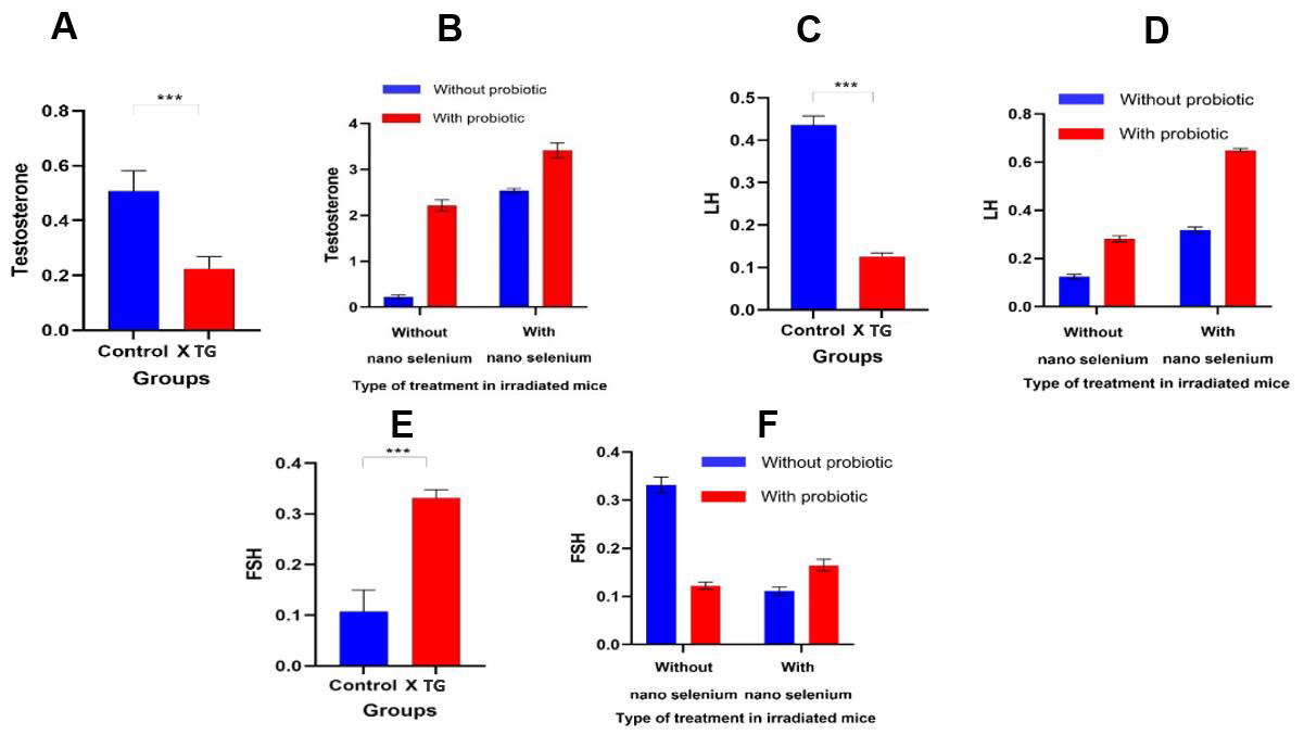
Figure 1. Means and standard deviations of: A&B – testosterone, C&D – luteinizing hormone (LH), and E&F – follicle-stimulating hormone (FSH) in control and irradiated Syrian mice, as well as in different treatment groups (TG).
Quantitative analysis revealed significant disruption of reproductive hormones following X-ray irradiation. Rodents in the irradiated group exhibited significant decreases in serum testosterone (P<0.0001) and LH (P<0.001). In contrast, FSH levels were significantly elevated in the X-ray-exposed group (P<0.001). Intervention with nano-selenium or probiotics significantly mitigated these deficits, resulting in a significant restoration of testosterone and LH levels (P<0.0001 for both treatments). Most importantly, the combined use of nano-selenium and probiotics exerted a synergistic effect, resulting in hormone levels exceeding those achieved with either treatment alone and approaching baseline control values (P<0.0001).
Both nano-selenium and probiotics were able to reduce FSH levels in mice exposed to X-rays (P<0.0001 and <0.001, respectively). However, FSH levels in animals receiving the combination of probiotics and nano-selenium were higher than in those receiving either compound alone, suggesting a possible antagonistic interaction between them in this regard.
Hematological parameters
Figure 2 shows the mean WBC, RBC, and platelet counts in different treatment groups.
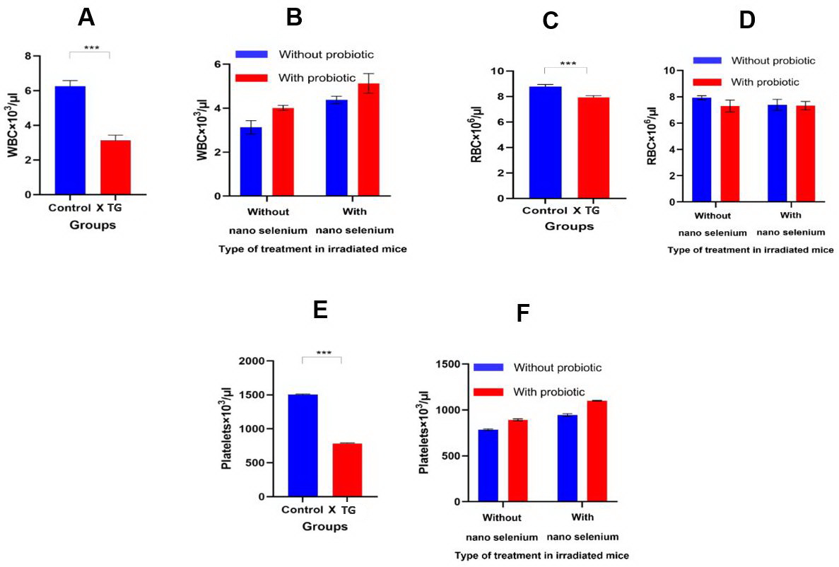
Figure 2. Means and standard deviations of: A&B – white blood cell (WBC), C&D – red blood cell (RBC), and E&F – platelet counts in control and irradiated Syrian mice, as well as in different treatment groups (TG).
X-ray irradiation resulted in significant reductions in WBC, RBC, and platelet counts vs. the control group (P<0.001, P<0.001, and P<0.0001, respectively). Monotherapy with nano-selenium or probiotics significantly attenuated X-ray-induced leukopenia (P<0.0001 and P<0.001, respectively) and thrombocytopenia (both P-values <0.0001). However, the combination of nano-selenium and probiotics did not demonstrate a synergistic effect on WBC count (P=0.506). Neither intervention significantly mitigated X-ray-induced anemia, as evidenced by the lack of recovery in RBC count.
Biochemical analyses
Mean values of biochemical parameters in different treatment groups are shown in Figure 3.
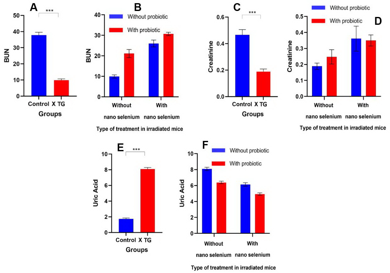
Figure 3. Means and standard deviations of: A&B – blood urea nitrogen (BUN), C&D – creatinine, and E&F – uric acid levels in control and irradiated Syrian mice, as well as in different treatment groups (TG).
Compared with the control group, X-ray irradiation significantly reduced BUN and creatinine levels but increased uric acid levels (all P-values <0.0001). Treatment with nano-selenium, probiotics, or a combination of both restored BUN and creatinine levels in irradiated mice.
Discussion
Radiation therapy remains a cornerstone of cancer treatment, but its undesirable side effects on radiosensitive organs such as the testes necessitate the development of effective radioprotection strategies. This study demonstrated that pretreatment with nano-selenium and L. casei, either individually or in combination, provides significant protection against X-ray-induced hormonal, hematological, and biochemical abnormalities in male Syrian mice. However, the protective effects were not uniform across all parameters, providing important mechanistic insights into the differential radiosensitivity of various cell lineages and the different pathways affected by these interventions.
The most striking result was the synergistic effect of combination treatment with nano-selenium and probiotics on testosterone and LH levels, which approached baseline control values, while FSH exhibited just an additive effect. This differential synergy may be explained by the distinct but complementary mechanisms by which these agents modulate the hypothalamic-pituitary-gonadal axis. The lack of synergy in FSH reduction may reflect the predominant role of Sertoli cell-produced inhibin B in the downregulation of FSH. Radiation-induced Sertoli cell damage can impair inhibin production, and although both treatments provide some protection, neither fully restores this specialized Sertoli cell function within 30 days.
The observation that both nano-selenium and probiotics attenuated radiation-induced leukopenia and thrombocytopenia but failed to restore RBC counts, and that combination therapy demonstrated no synergy in restoring WBC counts, highlights differences in radiosensitivity and the kinetics of hematopoietic stem cell regeneration depending on their cell lineage. Hematopoietic stem cells and progenitor populations exhibit varying radiosensitivity depending on their proliferation rate and oxidative metabolism. Mechanistically, both nano-selenium and probiotics may preserve the bone marrow microenvironment that supports the maintenance and differentiation of certain cell lineages.
The significant reduction in BUN and creatinine levels after irradiation, followed by a recovery to control values with nano-selenium and probiotics, requires careful interpretation. This pattern likely indicates radiation-induced metabolic dysregulation, which normalizes after treatment, rather than nephrotoxicity. Irradiation can induce a catabolic state. This can lead to decreased protein synthesis and increased protein breakdown, which paradoxically can reduce BUN levels. The observed decrease in creatinine concentration may reflect radiation-induced muscle atrophy or decreased creatine metabolism. Thus, the return of BUN and creatinine levels to values close to control in the treatment groups indicates the maintenance of normal protein metabolism and muscle mass, which is consistent with the systemic radioprotective effect of these agents.
These beneficial effects may be achieved through modulation of inflammatory pathways, metabolic processes, and the oxidative stress pathway. Previous studies have demonstrated the advantageous effects of selenium on multiple organ systems. For example, selenium enhances reproductive function in animals such as mice, pigs, and sheep [12]. Furthermore, selenium treatment was found to protect mouse testes from the toxic effects of radiation on all clinical parameters except edema. For some factors, such as spermatogenesis arrest and seminiferous tubule atrophy, selenium can completely suppress radiation toxicity [26]. The effect of selenium on RBC viability has also been investigated. Selenium deficiency is a factor that suppresses glutathione peroxidase activity and increases the amount of oxygen free radicals in tissues and various cell types, including RBCs [27-29].
In a previous study, we demonstrated the radioprotective effect of nano-selenium and a probiotic in the same Syrian mice models, confirming their potent antioxidant activity and effects on increasing the mean number of spermatogonia, sperm count, and rates of spermatogenesis and sperm motility [23].
The mechanism of cell protection by radioprotectors is through the inhibition of free radicals. An ideal radioprotector should reduce the adverse effects of ionizing radiation with low tissue toxicity. For this reason, the use of substances naturally present in the body has attracted considerable attention in this field. Selenium is an essential nutrient found in both organic and inorganic forms. Adequate selenium levels enhance antioxidant activity and support overall health. Recently, selenium nanoparticles have attracted widespread attention due to their high availability and lower toxicity compared to organic and inorganic forms of selenium. It is worth noting that nano-selenium-enriched probiotics exhibit a number of beneficial effects due to the modulation of antioxidant properties and cytokine production [30]. It was revealed that the combination of probiotics and nano-selenium significantly improved the activity of intracellular antioxidant enzymes, viz., thioredoxin reductase, glutathione peroxidase, and glutathione reductase [30].
Consistent with our results, a previous study showed that cell-free extracts of Lactococcus lactis produced by ionizing radiation can significantly reduce 60Coγ-induced oxidative stress by increasing the activity of several antioxidant enzymes and reducing malondialdehyde levels in serum, liver, and spleen. This finding suggested that L. lactis is a natural radioprotective agent [31].
Conclusion
Our study delivers the first evidence that combined administration of nano-selenium and L. casei provides differential, lineage-specific radioprotection in male Syrian mice. Key novel findings include synergistic restoration of testosterone and LH levels through complementary effects on Leydig cell steroidogenesis (selenium) and hypothalamic GnRH regulation (probiotics); additive protection of leukocyte and platelet counts; and differential lineage-specific effects, with erythroid progenitors showing no significant restoration within 30 days post-irradiation, reflecting their unique dependence on additional signaling pathways not affected by this treatment. These results provide the basis for mechanism-based radioprotection strategies combining nano-selenium and probiotics. The specific molecular pathways involved in these effects need to be elucidated.
Future prospects
Despite these promising results, translating these data into clinical practice faces several challenges and opens up opportunities for future research. Determining the optimal therapeutic window for nano-selenium is crucial, as selenium has a narrow range between beneficial and toxic doses. Long-term biodistribution and elimination of nanoparticles from the body require careful study. Furthermore, standardizing probiotic preparations and ensuring their stability and functionality in the intestine after irradiation pose significant challenges. Finally, the precise interaction between the gut microbiome (modulated by probiotics) and systemic radioprotection, particularly in the context of the hormonal and hematopoietic systems, should be assessed in future studies.
Limitations
Several limitations should be considered when interpreting our results. First, we did not directly measure molecular endpoints. Second, the 30-day observation period after irradiation, although sufficient for one spermatogenetic cycle, may not encompass full recovery of hematopoiesis, particularly RBC counts. Longer time intervals (60-90 d) would allow us to clarify whether RBC counts will ultimately recover. Third, we used single doses of nano-selenium and L. casei. Dose-response studies are needed to determine the optimal therapeutic ranges and the feasibility of achieving erythroid protection at higher doses. Fourth, the use of non-inbred Syrian mice, although enhancing translational relevance, introduces genetic variability that may mask subtle treatment effects. Studies using inbred strains with a defined genetic background could confirm specific mechanisms. Fifth, we did not measure tissue selenium concentrations or test the colonization efficiency of L. casei. Direct measurement of selenium levels in target organs and quantification of fecal L. casei would confirm adequate delivery and colonization. Sixth, this study was conducted exclusively on males; the radioprotective effects on the female reproductive system remain unknown. Furthermore, we used a single acute dose of radiation rather than clinically relevant fractionated protocols, which may induce different biological responses.
Ethical approval and consent to participate
The authors followed the guidelines for the care and use of laboratory animals by the U.S. National Institutes of Health and confirmed compliance with these guidelines in the Materials and Methods section of this manuscript. Furthermore, the full study protocol was approved by the Ethics Committee of Tehran University of Medical Sciences (IR.TUMS.MEDICINE.REC.1400.1104) under grant no. 99-1-148-46308.
Availability of data and materials
All data obtained or analyzed during this study are presented in this published article.
Conflict of interest
The authors declare no conflicts of interest.
Funding
No external funding was received for the study.
Author contributions
AE performed the experiments. FK, SK, and MT collected and analyzed the data. QT, MHM, and EM supervised the study. SGF edited the manuscript. All authors approved the final version of the article.
Statement on the use of artificial intelligence
No AI tools were used in generating the results, figures, or text, revising the manuscript, compiling the references, or for any other purpose.
- Weiss JF, Landauer MR. History and development of radiation-protective agents. Int J Radiat Biol 2009; 85(7): 539-573. https://doi.org/10.1080/09553000902985144.
- Guo C, Wang Q, Shuai P, Wang T, Wu W, Li Y, et al. Radiation and male reproductive system: Damage and protection. Chemosphere 2024; 357: 142030. https://doi.org/10.1016/j.chemosphere.2024.142030.
- Meistrich ML. Effects of chemotherapy and radiotherapy on spermatogenesis in humans. Fertil Steril 2013; 100(5): 1180-1186. https://doi.org/10.1016/j.fertnstert.2013.08.010.
- Okano T, Ishiniwa H, Onuma M, Shindo J, Yokohata Y, Tamaoki M. Effects of environmental radiation on testes and spermatogenesis in wild large Japanese field mice (Apodemus speciosus) from Fukushima. Sci Rep 2016; 6: 23601. https://doi.org/10.1038/srep23601.
- Simon L, Proutski I, Stevenson M, Jennings D, McManus J, Lutton D, et al. Sperm DNA damage has a negative association with live-birth rates after IVF. Reprod Biomed Online 2013; 26(1): 68-78. https://doi.org/10.1016/j.rbmo.2012.09.019.
- Gharagozloo P, Gutiérrez-Adán A, Champroux A, Noblanc A, Kocer A, Calle A, et al. A novel antioxidant formulation designed to treat male infertility associated with oxidative stress: promising preclinical evidence from animal models. Hum Reprod 2016; 31(2): 252-262. https://doi.org/10.1093/humrep/dev302.
- Citrin D, Cotrim AP, Hyodo F, Baum BJ, Krishna MC, Mitchell JB. Radioprotectors and mitigators of radiation-induced normal tissue injury. Oncologist 2010; 15(4): 360-371. https://doi.org/10.1634/theoncologist.2009-S104.
- Raviraj J, Bokkasam VK, Kumar VS, Reddy US, Suman V. Radiosensitizers, radioprotectors, and radiation mitigators. Indian J Dent Res 2014; 25(1): 83-90. https://doi.org/10.4103/0970-9290.131142.
- Mileikowsky C. Radiation therapy for cancer patients. Patent Number: 4,705,955. Date of Patent: Nov. 10, 1987. https://patents.google.com/patent/US4705955A/en.
- Daniels LA. Selenium metabolism and bioavailability. Biol Trace Elem Res 1996; 54(3): 185-199. https://doi.org/10.1007/BF02784430.
- Liu C, Chen Y, Zhang J, Huang M, Su Q, Lu Z, et al. Preliminary studies on influence of selenium deficiency to the developments of genital organs and spermatogenesis of infancy boars. Acta Vet Zootech Sin 1982; 13: 73-77.
- Combs GF Jr, Combs SB, The role of selenium in nutrition. Academic Press, Inc. 1986; 544 p. https://doi.org/10.1016/B978-0-12-183495-1.X5001-5.
- Berger MM. Can oxidative damage be treated nutritionally? Clin Nutr 2005; 24(2): 172-183. https://doi.org/10.1016/j.clnu.2004.10.003.
- Hoffmann PR, Berry MJ. The influence of selenium on immune responses. Mol Nutr Food Res 2008; 52(11): 1273-1280. https://doi.org/10.1002/mnfr.200700330.
- Forootanfar H, Adeli-Sardou M, Nikkhoo M, Mehrabani M, Amir-Heidari B, Shahverdi AR, et al. Antioxidant and cytotoxic effect of biologically synthesized selenium nanoparticles in comparison to selenium dioxide. J Trace Elem Med Biol 2014; 28(1): 75-79. https://doi.org/10.1016/j.jtemb.2013.07.005.
- Khurana A, Tekula S, Saifi MA, Venkatesh P, Godugu C. Therapeutic applications of selenium nanoparticles. Biomed Pharmacother 2019; 111: 802-812. https://doi.org/10.1016/j.biopha.2018.12.146.
- Lavanya M, Namasivayam SKR, John A. Developmental Formulation Principles of Food Preservatives by Nanoencapsulation-Fundamentals, Application, and Challenges. Appl Biochem Biotechnol 2024; 196(10): 7503-7533. https://doi.org/10.1007/s12010-024-04943-1.
- Sowmya R, Namasivayam SKR, Sivasuriyan KS, Varshan GSA. Nano-selenium-Derived Polymeric Bionanocomposites Induced Antibacterial, Anti-biofouling Efficacy Against Enterobacterial Waterborne Pathogens, and Their Ecotoxicity Assessment. BioNanoSci 2025; 15: 107. https://doi.org/10.1007/s12668-024-01667-8.
- Dinan TG, Quigley EM. Probiotics in the treatment of depression: science or science fiction? Aust N Z J Psychiatry 2011; 45(12): 1023-1025. https://doi.org/10.3109/00048674.2011.613766.
- Cats A, Kuipers EJ, Bosschaert MA, Pot RG, Vandenbroucke-Grauls CM, Kusters JG. Effect of frequent consumption of a Lactobacillus casei-containing milk drink in Helicobacter pylori-colonized subjects. Aliment Pharmacol Ther 2003; 17(3): 429-435. https://doi.org/10.1046/j.1365-2036.2003.01452.x.
- Adams CA. The probiotic paradox: live and dead cells are biological response modifiers. Nutr Res Rev 2010; 23(1): 37-46. https://doi.org/10.1017/S0954422410000090.
- Yue Y, Xu X, Yang B, Lu J, Zhang S, Liu L, et al. Stable Colonization of Orally Administered Lactobacillus casei SY13 Alters the Gut Microbiota. Biomed Res Int 2020; 2020: 5281639. https://doi.org/10.1155/2020/5281639.
- Ehghaghi A, Zokaei E, Modarressi MH, Tavoosidana G, Ghafouri-Fard S, Khanali F, et al. Antioxidant and anti-apoptotic effects of selenium nanoparticles and Lactobacillus casei on mice testis after X-ray. Andrologia 2022; 54(11): e14591. https://doi.org/10.1111/and.14591.
- El-Ghazaly MA, Fadel N, Rashed E, El-Batal A, Kenawy SA. Anti-inflammatory effect of selenium nanoparticles on the inflammation induced in irradiated rats. Can J Physiol Pharmacol 2017; 95(2): 101-110. https://doi.org/10.1139/cjpp-2016-0183.
- Norouzi R, Mirzaei F, Siadatpanah A, Adnani Sadati SJ. Evaluation of anti-leishmanial effect of selenium nanoparticles on Leishmania major promastigotes in vitro. Avicenna J Clin Microbiol Infect 2022; 9(3): 119-123. https://doi.org/10.34172/ajcmi.2022.3393.
- Bagheri H, Salajegheh A, Javadi A, Amini P, Shekarchi B, Shabeeb D, et al. Radioprotective Effects of Zinc and Selenium on Mice Spermatogenesis. J Biomed Phys Eng 2020; 10(6): 707-712. https://doi.org/10.31661/jbpe.v0i0.957.
- Van Vleet JF. Comparative efficacy of five supplementation procedures to control selenium-vitamin E deficiency in swine. Am J Vet Res 1982; 43(7): 1180-1189. https://pubmed.ncbi.nlm.nih.gov/6213185.
- Fraga CG, Arias RF, Llesuy SF, Koch OR, Boveris A. Effect of vitamin E- and selenium-deficiency on rat liver chemiluminescence. Biochem J 1987; 242(2): 383-386. https://doi.org/10.1042/bj2420383.
- Lessard M, Yang WC, Elliott GS, Rebar AH, Van Vleet JF, Deslauriers N, et al. Cellular immune responses in pigs fed a vitamin E- and selenium-deficient diet. J Anim Sci 1991; 69(4): 1575-1582. https://doi.org/10.2527/1991.6941575x.
- Liu R, Sun W, Sun T, Zhang W, Nan Y, Zhang Z, et al. Nano selenium-enriched probiotic Lactobacillus enhances alum adjuvanticity and promotes antigen-specific systemic and mucosal immunity. Front Immunol. 2023; 14: 1116223. https://doi.org/10.3389/fimmu.2023.1116223.
- Suo K, Chen S, Li X, Liu X, Yi J, Zhu J, et al. Radioprotective effect of radiation-induced Lactococcus lactis cell-free extract against 60Coγ injury in mice. J Dairy Sci 2021; 104(9): 9532-9542. https://doi.org/10.3168/jds.2021-20291.
Received 8 December 2025, Revised 27 April 2026, Accepted 6 June 2026
© 2025, Russian Open Medical Journal
Correspondence to Soudeh Ghafouri-Fard & Elahe Motevaseli. E-mails: s.ghafourifard@sbmu.ac.ir, e_motevaseli@tums.ac.ir.