

Introduction
Lung cancer is one of the most common and deadly cancers worldwide. It is classified into two main types: non-small cell lung cancer (NSCLC) and small cell lung cancer (SCLC). NSCLC accounts for more than 80-85% of cases, while SCLC accounts for approximately 15-20% [1, 2]. Many patients are diagnosed with lung cancer at an advanced stage, which complicates treatment.
Available treatment methods include surgery, chemotherapy, radiation therapy, and targeted therapy [3]. Pemetrexed (PEM) is a chemotherapy drug developed for the treatment of NSCLC and certain other cancers. It acts as an antimetabolite, inhibiting cancer cell proliferation by blocking enzymes critical for DNA and RNA production, particularly dihydrofolate reductase (DHFR) and thymidylate synthase (TYMS) [4]. These enzymes play an important role in folate metabolism and nucleotide synthesis. By inhibiting them, PEM limits the resources required by cancer cells for growth and division, thereby promoting tumor shrinkage and disease control [4, 5]. Limitations of chemotherapy drugs include damage to healthy cells and serious side effects, low tumor permeability, and cancer cell resistance to drugs [6].
Nanoparticles (NPs) have significant potential to enhance the efficacy of chemotherapeutic agents by improving drug permeability and targeted delivery, and can also reduce side effects and facilitate targeted cancer treatment by increasing the formation of reactive oxygen species (ROS) and stimulating tumor cell apoptosis [7]. Copper oxide (CuO) NPs exhibit anticancer properties due to their antioxidant capabilities and ROS formation. As a result, cancer cell growth becomes inhibited. CuO NPs induce oxidative stress, leading to cell death, and can also function as targeted drug carriers, enhancing the efficacy of chemotherapy and reducing side effects [8]. Furthermore, studies show that CuO NPs show higher cytotoxicity towards human cells than to other metal oxide NPs. The anticancer effects of CuO NPs have been investigated in various types of cancer, including liver, lung, breast, cervical, and pancreatic cancer [9].
MicroRNAs (miRNAs) are non-coding RNAs that regulate gene expression by binding to messenger RNAs (mRNAs), influencing biological processes such as cell division, death, and migration [10]. Studies show that abnormal miRNA expression significantly contributes to the development and progression of various metabolic diseases. In cancer, altered miRNA levels can influence the expression of genes associated with cancer cell growth, with some functioning as oncogenes and others as tumor suppressors [11]. The miR-548 gene family, comprising 69 poorly conserved primate-specific genes on human chromosomes, is critical for a variety of diseases. It generates mature miR-548c-3p, which has been identified as a regulatory factor in many cancers, including prostate, esophageal, thyroid, osteosarcoma, glioma, gallbladder, gastric, and breast cancers [12]. Furthermore, mounting evidence suggests that miR-548 inhibits cancer cell proliferation and promotes apoptosis, acting as a tumor suppressor in the lung [13]. The present study aimed to investigate the antitumor effects of CuO NPs co-administered with PEM and to examine changes in hsa-miR-548c-3p expression in lung cancer cells.
Material and Methods
Experimental design
A549 cells (ATCC® CCL185™) were obtained from the Pasteur Institute of Iran and cultured in DMEM (Sigma, St. Louis, MO, USA) with 10% FBS at 37 °C in a 5% CO2 atmosphere. Cells were exposed to different concentrations of PEM (1–12 μM) [14] and CuO NPs (Nanosany-Iran) (0, 2.5, 5, 10, 20, 40, 80, 100, 200, and 400 μg/mL) [15] for 24 and 48 h to determine the IC50 concentration. Afterwards, the cells were divided into four groups: control, CuO NPs-treated group, PEM-treated group, and combination therapy group receiving both CuO NPs and PEM. For treatment in the combination therapy group (CuO NPs + PEM), half the IC50 concentration was used.
Cell viability
After 48 h of treatment of A549 cells with CuO NPs, PEM, and a combination of CuO NPs and PEM, the culture medium was carefully removed from each well. Fresh culture medium (90 µL) was added to each well along with 10 µL of a 5 mg/mL MTT solution (resulting in a final concentration of 0.5 mg/mL). The plates were incubated for 4 h, and formazan crystal formation was observed under an inverted microscope. The medium was carefully removed, and 100 µL of DMSO (Sigma Aldrich, USA) was added to each well. After incubation for 30 minutes in order to dissolve the crystals, the resulting purple color was measured with an ELISA reader at a wavelength of 570 nm. All concentrations were tested in triplicate to ensure precision [14].
Apoptosis assay
We used fluorescent dyes, specifically acridine orange and ethidium bromide, to stain cell nuclei. Acridine orange can penetrate the membranes of living cells, while ethidium bromide only penetrates the membranes of dead cells. In this assay, living cells exhibit green fluorescence, while apoptotic cells change color from orange to red. A suspension of 104 cells in DMEM was placed in 24-well plates (500 µL per well) and incubated for 24 h. Then the wells were processed according to the experimental design. After 48 h, the treatment solutions were removed, and a mixture (250 µL) containing acridine orange and ethidium bromide (5 µg/mL of the former and 5 µg/mL of the latter in phosphate-buffered saline) were added for 2 min. Cells were subsequently examined by fluorescence microscopy [14].
LDH release assay
Lactate dehydrogenase (LDH) is an enzyme that serves as a marker of cell damage, and cytotoxicity assays rely on its release. This enzyme is present in eukaryotic cells and is released from the plasma membrane upon cell damage or death. We assessed LDH levels using the Kiazist kit (KLDH96): 10,000 cells were seeded into each well of a 96-well plate and incubated for 48 h. After that, treatment groups were applied, and 20 µL of Permi solution was added to each well. The plate was then incubated at room temperature for 1 h. To measure LDH, 50 µL from each well was transferred to a new plate, after which working buffer was added (50 µL). After 30 minutes of incubation at 37 °C in the dark, absorbance was measured at 570 nm [14].
ROS production assay
ROS are reactive oxygen-containing molecules that are produced naturally in mitochondria or by external agents in cells. These radicals can damage cellular macromolecules, causing various tissue and cellular disorders, including cancer. Common types of ROS include superoxide radicals, hydroxyl radicals, and peroxides. In this assay, the 2',7'-dichlorofluorescein diacetate (DCFDA) dye was employed to measure ROS levels. This dye can penetrate living cells and be de-esterified by intracellular enzymes, after which it fluoresces upon reduction by specific ROS. Cells were seeded in a 96-well plate, treated accordingly, and then washed with ROS buffer. Afterwards, 100 µL of DCFDA working solution was added to each well and incubated in the dark at 37 °C for 45 min. Fluorescence intensity was then measured at wavelengths of 485/528 nm [14].
RNA extraction
RNA extraction from cell cultures was performed using the RNX Plus kit. First, 1 mL of the RNX Plus solution was mixed with 106 cells and transferred to a microtube. Next, 250 μL of cold chloroform was added, the mixture was vortexed, and then centrifuged for 15 min at 13,000 rpm at 4 °C. The resulting clear supernatant was carefully transferred to a new microtube, and 500 μL of isopropanol was added. This mixture was then centrifuged for another 15 min at 13,000 rpm at 4 °C, resulting in the formation of a white precipitate indicating the presence of RNA. Following this, 1 mL of 75% ethanol was added, and the sample was centrifuged for 10 min at 8,500 rpm at 4 °C. The supernatant was drained, and after the precipitate had dried, 50 μL of RNase-free water treated with diethyl pyrocarbonate (DEPC) was added. The quality and quantity of the isolated RNA were assessed using a NanoDrop spectrophotometer, after which the RNA was stored at -70 °C [14].
cDNA synthesis
The AddScript cDNA Synthesis Kit was used for cDNA synthesis. In this process, 1 μg of DNase I-treated RNA was placed in a 0.2 mL microtube. To it were added 1 μL of reverse transcriptase enzyme, 2 μL of dNTPs, 4 μL of buffer, and 3 μL of a mixture of oligo(dT) and random hexamer primers. The total volume was adjusted to 20 μL with DEPC-treated water. The mixture was then incubated for 60 min at 50 °C, followed by a 10-min incubation at 80 °C. The synthesized cDNA was stored at -21 °C until real-time PCR was performed. For miR cDNA synthesis, a specific primer was used instead of oligo(dT) and random hexamer primers, as detailed in Table 1 [14].
Table 1. Hsa-miR-548c-3p and U48 sequences, and primers used for real-time qRT-PCR
|
miRNA |
Sequence |
Primer sequence |
|
|
hsa-miR-548c-3p (MIMAT0003285) |
CAAAAAUCUCAAUUACUUUUGC |
Stem-loop primer: GTCGTATCCAGTGCAGGGTCCGAGGTATTCGCACTGGATACGACGCAAAAG Forward primer: GCACAGCCAAAAATCTCAATTAC Reverse primer: GTCGTATCCAGTGCAGGGT |
|
|
U48 (NR_002745.1) |
AGTGATGATGACCCCAGGTAACTCTTGAGTGTGTCGCTGATGCCATCACCGCAGCGCTCTGACC |
Stem-loop primer: GTCGTATCCAGTGCAGGGTCCGAGGTATTCGCACTGGATACGACGGTCAG Forward primer: CTCTGAGTGTGTCGCTGATGCC Reverse primer: CCAGTGCAGGGTCCGAGGTA |
|
qRT-PCR, real-time quantitative reverse transcription polymerase chain reaction.
Real-time PCR
To assess changes in gene expression, we performed the SYBR Green-based real-time PCR according to the guidelines provided with the Parstus kit. First, 10 µL of the SYBR Green master mix was placed in a real-time PCR microtube. Then, 1 µL of a mixture containing the forward and reverse primers for the target gene, each at a concentration of 10 µM (as indicated in Tables 1 and 2), was added. Next, 1 µL of the prepared cDNA was added, and the final volume was adjusted to 20 µL with distilled water [14].
Table 2. Primer sequences, annealing temperatures, and PCR product sizes shown in the (5′→3′) direction
|
Gene |
|
Primer sequence (5′→3′) |
Length (bp) |
Annealing temperature (°C) |
Product size (bp) |
|
TYMS(NM_001071.4) |
F |
CTGGGGCAGATCCAACACAT |
20 |
60 |
152 |
|
R |
ACACCCTTCCAGAACACACG |
20 |
|||
|
CASP8(NM_001228.5) |
F |
CGGGGATACTGTCTGATCAT |
20 |
57 |
127 |
|
R |
CAAAGGTCGTGGTCAAAGC |
19 |
|||
|
CASP9(NM_001229.5) |
F |
TGGCTCCTGGTACGTTGA |
18 |
57 |
99 |
|
R |
GAAACAGCATTAGCGACCC |
19 |
|||
|
GAPDH(NM_002046.7) |
F |
TGACTTCAACAGCGACACCCA |
21 |
61 |
121 |
|
R |
CACCCTGTTGCTGTAGCCAAA |
21 |
Statistical analysis
Statistical analysis and plotting of cell data were performed using GraphPad Prism, version 8.2.1. One-way analysis of variance (ANOVA) was employed to compare different groups. For all statistical evaluations, P<0.05 was considered statistically significant, and data are presented as mean ± standard error of the mean (SEM). Relative gene expression levels were estimated using the Relative Expression Software Tool (REST), version 2.0.13. Results obtained using REST were analyzed alongside cell data to ensure a thorough and comprehensive analysis.
Results
IC50 for PEM and CuO NPs
IC50 concentrations for PEM and CuO NPs were determined using the MTT assay. IC50 measures the potency of a substance in terms of inhibiting a specific biological function: it indicates the amount required to achieve 50% inhibition of a biological process or component in vitro. Table 3 shows that the concentrations of PEM and CuO NPs required to achieve 50% inhibition of A549 cell growth after 24 h were 4.766 μM and 160.2 μg/mL, respectively, and after 48 h, 2.891 μM and 72.12 μg/mL.
Table 3. Percentage viability and IC50 calculation for A549 cells after 24 and 48 h of treatment with PEM and CuO NPs
|
Group |
IC50 24 hours |
IC50 48 hours |
|
PEM |
4.766 µM |
2.891 µM |
|
CuO NPs |
160.2 µg/ml |
72.12 µg/ml |
Cell viability
The greatest reduction in cancer cell viability was observed after combination therapy with PEM and CuO NPs. Figure 1 shows that all treatments (CuO NPs, PEM, and their combination) statistically significantly reduced cell viability vs. the control group (P<0.05). Notably, the group receiving combination therapy (CuO NPs + PEM) experienced the most pronounced reduction in viability (P<0.05).
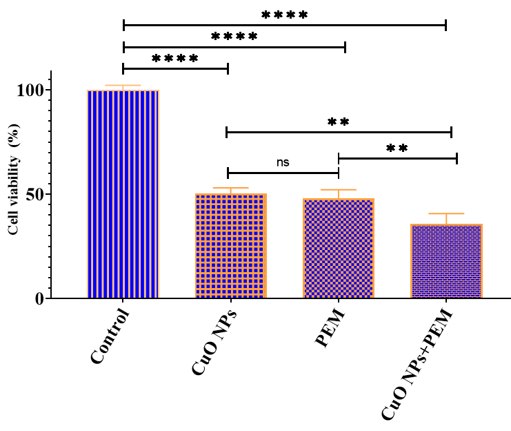
Figure 1. Post-treatment comparison of A549 cell viability with the study groups.
**, P<0.01; ****, P<0.0001; ns, P>0.05.
Apoptosis
The percentage of apoptosis in cancer cells after combination therapy with PEM and CuO NPs showed the most significant increase. Figure 2 illustrates the levels of apoptosis in the different treatment groups. The control group (no treatment) had the lowest levels of apoptosis, as no external factors were applied. In contrast, both PEM and CuO NPs significantly increased apoptosis, demonstrating a significant difference from the control group (P<0.0001). The combination therapy group (CuO NPs + PEM) exhibited the highest percentage of apoptosis (P<0.0001).
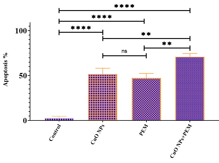
Figure 2. Post-treatment comparison of apoptosis in A549 cells with the study groups.
**, P<0.01; ****, P<0.0001; ns, P>0.05.
ROS
As shown in Figure 3, ROS formation significantly increased in all treatment groups vs. the control group (P<0.05). Furthermore, a significant increase in ROS production was observed in the combination therapy group (CuO NPs + PEM) vs. either monotherapy group (P<0.0001).
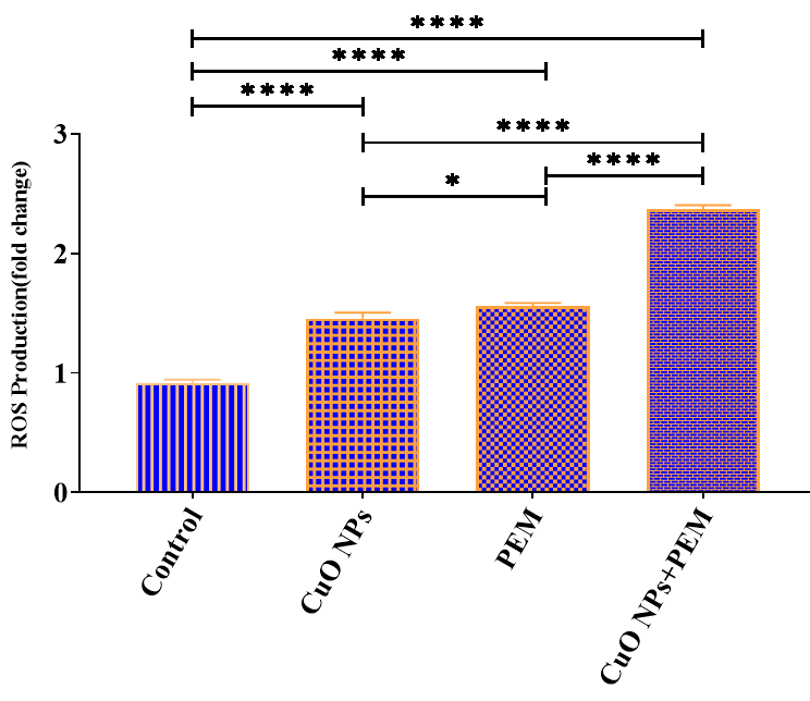
Figure 3. Post-treatment comparison of ROS formation in A549 cells with the study groups.
*, P<0.05; **, P<0.01; ****, P<0.0001; ns, P>0.05.
LDH
Figure 4 shows that LDH release levels increased significantly in all treatment groups vs. the control group (P<0.05). Furthermore, the combination therapy group (CuO NPs + PEM) demonstrated a highly significant increase in LDH release vs. either monotherapy group (P<0.0001).
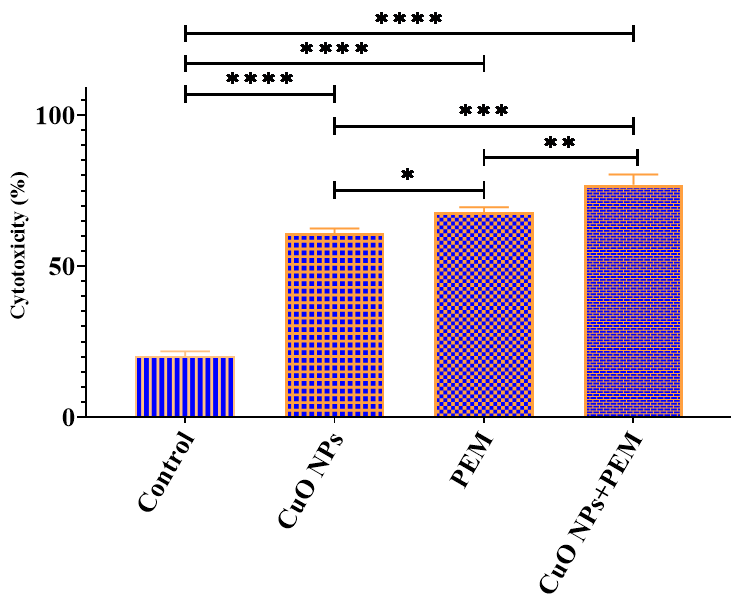
Figure 4. Post-treatment comparison of LDH release by A549 cells with the study groups.
*, P<0.05; **, P<0.01; ***, P<0.001; ****, P<0.0001; ns, P>0.05.
Real‑time PCR
The relative expression levels of hsa-miR-548c-3p, CASP8, and CASP9 in cancer cells increased after all treatments compared with untreated cells, with a more significant increase in the combination therapy group than in the other groups (P<0.0001). In contrast, the relative expression of TYMS in cancer cells decreased after all treatments, and this reduction was more pronounced in the combination therapy group than in the other groups (Figure 5).
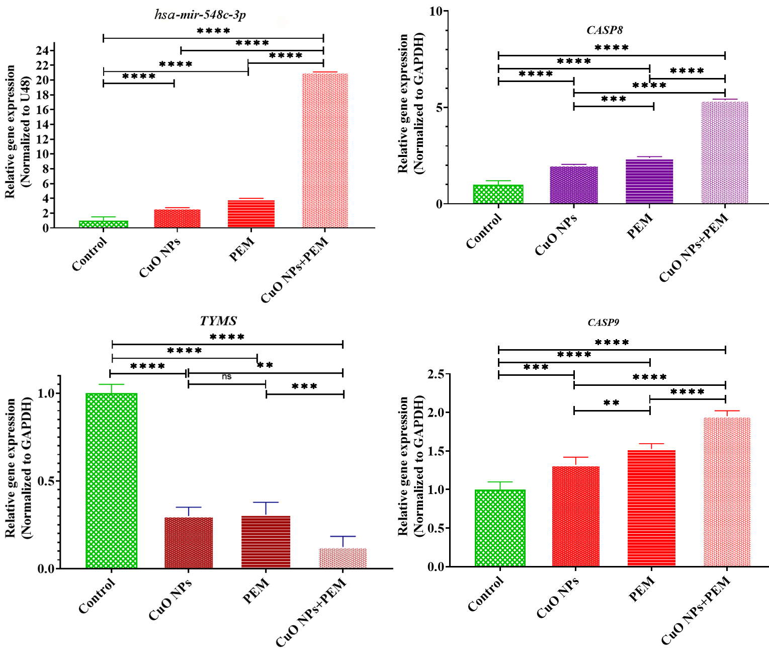
Figure 5. Relative expression of hsa-miR-548c-3p, CASP8, CASP9, and TYMS. Relative expression of hsa-miR-548c-3p in treated cells compared with untreated cells, normalized to RNU48. Relative expression of TYMS, CASP8, and CASP9 in treated cells vs. untreated cells, normalized to GAPDH.
*, P<0.05; ns, P>0.05.
Discussion
Our study examined the combined effect of CuO NPs on enhancing the cytotoxic efficacy of PEM in human lung cancer cells, specifically the A549 cell line. The results showed that the use of PEM in conjunction with CuO NPs significantly reduced cell viability, increased the percentage of apoptosis, enhanced ROS production, and resulted in higher LDH release compared with the control group and groups treated with either agent alone.
Enhanced apoptosis in cancer cells is considered a beneficial indicator for cancer therapy, as apoptosis is a natural and regulated mechanism that promotes the removal of damaged and abnormal cells. This process plays a crucial role in maintaining cellular homeostasis and preventing the proliferation of potentially harmful cells [16]. Moreover, ROS formation is recognized as a critical mechanism in the induction of apoptosis. ROS can trigger various signaling pathways that lead to programmed cell death, thereby promoting the elimination of cancer cells and increasing the effectiveness of anticancer treatment [17]. When cancer cell membranes are damaged in cell culture, LDH is released into the culture medium. This process is recognized as a marker of cell damage and is widely used in toxicology studies and to assess the therapeutic effect on cancer cells. LDH release following membrane damage indicates cell death or severe cell damage [18].
Our findings confirmed that CuO NPs may act as a potentiating agent in PEM therapy, promoting apoptotic responses in cancer cells. Furthermore, the results showed that combination therapy resulted in increased expression of hsa-miR-548c-3p, as well as increased expression of CASP8 and CASP9, while simultaneously diminishing TYMS expression. MiR-548c-3p reduces lung cancer cell proliferation by interfering with the galectin-3-mediated TLR4 signaling pathway. Furthermore, it increases the sensitivity of colorectal cancer cells to 5-fluorouracil by downregulating TYMS and ABCG2 [13, 19].
High TYMS expression in malignant tumors is associated with reduced sensitivity to antifolate drugs, including PEM. In particular, TYMS are recognized as important enzymes involved in folate metabolism and nucleotide synthesis. A decrease in their activity may lead to inhibition of cancer cell growth and proliferation [20]. Takezawa et al. (2011) showed that NSCLC cells overexpressing TYMS exhibited significantly reduced sensitivity to the antiproliferative effect of PEM, compared with control cells. TYMS overexpression reduced the inhibition of DNA synthesis and the induction of apoptosis by PEM. Furthermore, tumors from TYMS-overexpressing cells in nude mice were resistant to PEM, indicating a negative correlation between TYMS levels and treatment response [20].
Shafagh et al. (2015) found that CuO NPs selectively target cancer cells without affecting normal cells, as demonstrated by the MTT assay. Their cytotoxic effect on K562 cells was associated with ROS production, apoptosis induction confirmed by staining, and increased P53 expression, indicating a mitochondria-mediated apoptotic pathway [21]. Caspases play a crucial role in apoptosis. Caspase-8 is involved in the extrinsic pathway, while caspase-9 initiates the intrinsic apoptotic pathway [22]. The results of our study showed that CuO NPs and PEM induce apoptosis in lung cancer cells through both intracellular and extracellular pathways. Esmaeili Gouvarchin Ghaleh et al. (2019) reported a significant decrease in cell viability and an increase in apoptosis in MCF7 cells when treated with CuO NPs in combination with radiotherapy and hyperthermia vs. the control group. This treatment also resulted in increased ROS formation, LDH levels, and a significant increase in caspase-3 and caspase-9 activity, while caspase-8 activity remained unchanged [23].
Ghorbani Alvanegh et al. (2024) found that PEM treatment significantly increased LDH release after 24 h in Calu-6 cells. Treated samples showed increased levels of hsa-miR-320a-3p and BAX, while VDAC1, STAT3, DHFR, and BCL2 expression decreased compared to control samples [24]. Furthermore, the researchers demonstrated that PEM reduced cell viability and increased apoptosis. After 48 hours, ROS production was higher than in control cells, and LDH release increased. Moreover, PEM significantly increased the expression of hsa-miR-320a, showing an approximately 12-fold increase [14].
Khalili et al. (2023) found that miR-548c-3p sensitized colorectal cancer cells to the chemotherapeutic drug 5FU by inhibiting the expression of TYMS and ABCG2 genes [19]. Shi et al. (2015) showed that miR-548-3p was downregulated in breast cancer, and its overexpression inhibited cell proliferation while promoting apoptosis. ECHS1 expression was increased in breast cancer tissues and was identified as a target of miR-548-3p. These results suggest the potential of miR-548-3p as a therapeutic target by regulating ECHS1 expression [25]. Yao et al. (2020) reported that both miR-423 and miR-548c-3p exhibited reduced expression in osteosarcoma. Increased levels of miR-432 and miR-548c-3p in osteosarcoma cells were found to inhibit tumor cell proliferation and enhance apoptosis. These miRNAs may function as tumor suppressors in osteosarcoma, and their expression levels may serve as important indicators for assessing the benign or malignant nature of the disease [26].
Conclusion
This study revealed that the combination of PEM with CuO NPs significantly affects cellular responses, resulting in decreased survival and increased apoptosis in treated cells. Increased ROS production and LDH release indicate a cytotoxic effect of this combination therapy. Furthermore, increased levels of pro-apoptotic markers such as hsa-miR-548c-3p, CASP8, and CASP9, along with decreased levels of metabolic enzymes such as TYMS, suggest a shift in cellular pathways that may enhance the efficacy of PEM. These results highlighted the potential of this combination strategy to improve cancer treatment outcomes, which warrants further examination of its mechanisms and clinical application. Limitations of the study include the lack of in vivo validation, which may impact the generalizability of the results. Furthermore, focusing solely on specific biomarkers limits understanding of broader molecular mechanisms. The potential long-term effects and safety of CuO NPs have not been thoroughly investigated.
Acknowledgements
This study is part of Ms. Sepideh Mokabberi’s PhD thesis. The authors express their gratitude to everyone who assisted us in this research.
Author contributions
SM, NB, HEGG, and GF developed the theoretical formalism, performed the analytical calculations, and conducted the numerical modeling. The authors contributed to the final version of the manuscript. NB supervised the project. Zigap AI was used to check this article for plagiarism.
Funding
The authors declare that no external funds, grants, or other support were received during the preparation of this manuscript.
Data availability
The datasets generated and/or analyzed during this study are available from the corresponding author upon reasonable request.
Ethical approval and consent to participate
The Ethics Committee of Baqiyatallah University of Medical Sciences approved the protocols of this study (IR.BPUMS.REC.1402.306).
Conflict of interest
The authors declare no conflicts of interest.
- Padinharayil H, Varghese J, John MC, Rajanikant GK, Wilson CM, Al-Yozbaki M, et al. Non-small cell lung carcinoma (NSCLC): Implications on molecular pathology and advances in early diagnostics and therapeutics. Genes Dis 2023; 10(3): 960-989. https://https://doi.org/10.1016/j.gendis.2022.07.023.
- Hendriks LEL, Remon J, Faivre-Finn C, Garassino MC, Heymach JV, Kerr KM, et al. Non-small-cell lung cancer. Nat Rev Dis Primers 2024; 10(1): 71. https://doi.org/10.1038/s41572-024-00551-9.
- Tang FH, Wong HYT, Tsang PSW, Yau M, Tam SY, Law L, et al. Recent advancements in lung cancer research: A narrative review. Transl Lung Cancer Res 2025; 14(3): 975-990. https://doi.org/10.21037/tlcr-24-979.
- Komiya T, Memmott RM, Blumenthal GM, Bernstein W, Ballas MS, De Chowdhury R, et al. A phase I/II study of pemetrexed with sirolimus in advanced, previously treated non-small cell lung cancer. Transl Lung Cancer Res 2019; 8(3): 247-257. https://doi.org/10.21037/tlcr.2019.04.19.
- Li T, Huang JB, Lu JG, Li R, Wang Y, Shi XR, et al. Plasma thymidylate synthase and dihydrofolate reductase mRNA levels as potential predictive biomarkers of pemetrexed sensitivity in patients with advanced non-small cell lung cancer. J Thorac Dis 2020; 12(12): 7313-7319. https://doi.org/10.21037/jtd-20-3185.
- Davodabadi F, Sajjadi SF, Sarhadi M, Mirghasemi S, Hezaveh MN, Khosravi S, et al. Cancer chemotherapy resistance: Mechanisms and recent breakthrough in targeted drug delivery. Eur J Pharmacol 2023; 958: 176013. https://doi.org/10.1016/j.ejphar.2023.176013.
- Sabir S, Thani ASB, Abbas Q. Nanotechnology in cancer treatment: revolutionizing strategies against drug resistance. Front Bioeng Biotechnol 2025; 13: 1548588. https://doi.org/10.3389/fbioe.2025.1548588.
- Abdolmaleki S, Aliabadi A, Khaksar S. Unveiling the promising anticancer effect of copper-based compounds: a comprehensive review. J Cancer Res Clin Oncol 2024; 150(4): 213. https://doi.org/10.1007/s00432-024-05641-5.
- Abdollahzadeh H, Pazhang Y, Zamani A, Sharafi Y. Green synthesis of copper oxide nanoparticles using walnut shell and their size dependent anticancer effects on breast and colorectal cancer cell lines. Sci Rep 2024; 14(1): 20323. https://doi.org/10.1038/s41598-024-71234-4.
- Nunes S, Bastos R, Marinho AI, Vieira R, Benício I, de Noronha MA, et al. Recent advances in the development and clinical application of miRNAs in infectious diseases. Noncoding RNA Res 2024; 10: 41-54. https://doi.org/10.1016/j.ncrna.2024.09.005.
- Singh V, Sen A, Saini S, Dwivedi S, Agrawal R, Bansal A, et al. MicroRNA significance in cancer: An updated review on diagnostic, prognostic, and therapeutic perspectives. EJIFCC 2024; 35(4): 265-284. https://pubmed.ncbi.nlm.nih.gov/39810890.
- Tan PY, Wen LJ, Li HN, Chai SW. MiR-548c-3p inhibits the proliferation, migration and invasion of human breast cancer cell by targeting E2F3. Cytotechnology 2020; 72(5): 751-761. https://doi.org/10.1007/s10616-020-00418-3.
- Chen X, Chen Y, Hu Q, Chen X, Huang L. MiR-548c inhibits lung cancer cell proliferation through suppression of Galectin-3-mediated TLR4 signaling pathway. Int J Clin Exp Med 2018; 11(2): 581-592. https://e-century.us/files/ijcem/11/2/ijcem0055106.pdf.
- Ghorbani Alvanegh A, Esmaeili Gouvarchin Ghaleh H, Mohammad Ganji S. The Growth of A549 cell line is inhibited by pemetrexed through up-regulation of hsa-MiR-320a expression. Adv Biomed Res 2024; 13: 50. https://doi.org/10.4103/abr.abr_483_23.
- Bhavana S, Kusuma CG, Gubbiveeranna V, Sumachirayu CK, Ravikumar H, Nagaraju S. Green route synthesis of copper oxide nanoparticles using Vitex altissima [L] leaves extract and their potential anticancer activity against A549 cell lines and its apoptosis induction. Inorganic and Nano-Metal Chemistry 2024; 54(10): 1012-1025. https://doi.org/10.1080/24701556.2022.2081195.
- Mirzayans R. What is apoptosis and why is it inhibited by the most important tumor suppressor (p53)? Int J Mol Sci 2025; 26(21): 10505. https://doi.org/10.3390/ijms262110505.
- An X, Yu W, Liu J, Tang D, Yang L, Chen X. Oxidative cell death in cancer: Mechanisms and therapeutic opportunities. Cell Death Dis 2024; 15(8): 556. https://doi.org/10.1038/s41419-024-06939-5.
- Kari S, Subramanian K, Altomonte IA, Murugesan A, Yli-Harja O, Kandhavelu M. Programmed cell death detection methods: A systematic review and a categorical comparison. Apoptosis 2022; 27(7-8): 482-508. https://doi.org/10.1007/s10495-022-01735-y.
- Khalili E, Afgar A, Rajabpour A, Aghaee-Bakhtiari SH, Jamialahmadi K, Teimoori-Toolabi L. MiR-548c-3p through suppressing Tyms and Abcg2 increases the sensitivity of colorectal cancer cells to 5-fluorouracil. Heliyon 2023; 9(11): e21775. https://doi.org/10.1016/j.heliyon.2023.e21775.
- Takezawa K, Okamoto I, Okamoto W, Takeda M, Sakai K, Tsukioka S, et al. Thymidylate synthase as a determinant of pemetrexed sensitivity in non-small cell lung cancer. Br J Cancer 2011; 104(10): 1594-1601. https://doi.org/10.1038/bjc.2011.129.
- Shafagh M, Rahmani F, Delirezh N. CuO nanoparticles induce cytotoxicity and apoptosis in human K562 cancer cell line via mitochondrial pathway, through reactive oxygen species and P53. Iran J Basic Med Sci 2015; 18(10): 993-1000. https://pubmed.ncbi.nlm.nih.gov/26730334.
- Aral K, Aral CA, Kapila Y. The role of caspase-8, caspase-9, and apoptosis inducing factor in periodontal disease. J Periodontol 2019; 90(3): 288-294. https://doi.org/10.1002/JPER.17-0716.
- Esmaeili Govarchin Ghaleh H, Zarei L, Mansori Motlagh B, Jabbari N. Using CuO nanoparticles and hyperthermia in radiotherapy of MCF-7 cell line: synergistic effect in cancer therapy. Artif Cells Nanomed Biotechnol 2019; 47(1): 1396-1403. https://doi.org/10.1080/21691401.2019.1600529.
- Ghorbani Alvanegh A, Arpanaei A, Esmaeili Gouvarchin Ghaleh H, Mohammad Ganji S. MiR-320a upregulation contributes to the effectiveness of pemetrexed by inhibiting the growth and invasion of human lung cancer cell line (Calu-6). Mol Biol Rep 2024; 51(1): 310. https://doi.org/10.1007/s11033-024-09207-z.
- Shi Y, Qiu M, Wu Y, Hai L. MiR-548c-3p functions as an anti-oncogenic regulator in breast cancer. Biomed Pharmacother 2015; 75: 111-116. https://doi.org/10.1016/j.biopha.2015.07.027.
- Yao J, Tan W, Wu W, Ye R, Li Y, Chen Y. Effects of miR-432 and miR-548c-3p on the proliferation and invasion of osteosarcoma cells. J BUON 2020; 25(3): 1562-1568. https://pubmed.ncbi.nlm.nih.gov/32862605.
Received 26 August 2025, Revised 6 February 2026, Accepted 9 April 2026
© 2025, Russian Open Medical Journal
Correspondence to Hadi Esmaeili Gouvarchin Ghaleh. E-mail: h.smaili69@yahoo.com.