

Introduction
Heart failure (HF) remains a global health problem and represents the final stage of the cardiovascular continuum. The role of mitochondria in the pathogenesis of HF is an area of ongoing research. Studying mitochondrial function in patients with HF and coronary artery disease (CAD) is particularly important, as both conditions can impair mitochondrial function [1, 2]. It is noteworthy that 90% of the energy used by the contraction-relaxation cycle of cardiomyocytes comes from de novo synthesis of adenosine triphosphate in mitochondria [3, 4].
A key characteristic of mitochondria, distinguishing them from other intracellular organelles, is the presence of their own mitochondrial DNA (mtDNA) [5]. When released extracellularly, mtDNA acts as mitochondrial damage-associated molecular patterns (mtDAMPs), triggering inflammatory responses [6]. Previous studies demonstrated that mtDNA copy number increases in peripheral blood during cardiac surgery, particularly during coronary artery bypass grafting (CABG) with cardiopulmonary bypass, and returns baseline levels within 24-72 hours postoperatively [7, 8]. Furthermore, it was shown that elevated mtDNA copy number before CABG is associated with the development of postoperative atrial fibrillation [6, 9], which is probably due to the proinflammatory effect of mtDNA. Studies of changes in mtDNA copy number after CABG were limited to the first 24-72 hours; therefore, they did not examine how mtDNA copy number changes in the days following CABG, which is the goal of our study.
The development of complications after on-pump CABG is primarily associated with inflammation. Such complications include atrial fibrillation, postpericardiotomy syndrome, and infection. These conditions promote active release of mtDNA from cells and an increase in mtDNA copy number in plasma. At the same time, elevated levels of mtDAMPs after CABG lead to the progression of these complications and an aggravating postoperative course [10]. Confirmation of the association between increased mtDNA copy number in plasma and complications after cardiac surgery may serve as a basis for a more targeted study of this parameter.
The mechanisms of mtDNA release from cells into the extracellular space remain poorly understood. Passive mechanisms are believed to involve mtDNA release into the extracellular space following apoptosis or cell necrosis [11]. Passive mtDNA release may involve an increase in mtDNA copy number due to mechanical cell damage resulting from CABG surgery. Another mechanism for mtDNA release from cells may involve active release within extracellular vesicles or release as neutrophil extracellular traps [12]. This particular mechanism may likely be involved in changes in plasma mtDNA copy number at later stages after CABG and may be associated with the development of postoperative complications.
In addition to an increase in mtDNA copy number, an increase in cytochrome c may be indirect evidence of ongoing mitochondrial damage. Cytochrome c is a component of the mitochondrial respiratory chain that is released into the blood when mitochondria are damaged [13].
The goal of this study was to determine changes in blood mtDNA copy number in patients who underwent on-pump CABG from preoperative baseline to Day 10 postoperatively and to evaluate its association with postoperative complications.
Material and Methods
Study design
The study included 26 patients scheduled for CABG with a median age of 67 years (interquartile range [IQR]: 58-71 years). This was a single-center, prospective, unblinded cohort study (protocol registered at ClinicalTrials.gov: NCT05770349). Inclusion criteria were as follows: presence of HF with left ventricular ejection fraction (LVEF) <50%; atherosclerotic plaque of 70% or more in at least two major coronary arteries; decision by the cardiac team to perform on-pump CABG; signed informed consent; and availability of biomaterial specimens. Exclusion criteria for the study were as follows: refusal of revascularization or participation; need for additional cardiac surgeries besides CABG; active cancer; presence of implanted devices; severe renal dysfunction (CKD-EPI estimated glomerular filtration rate <30 mL/min/1.73 m2); infiltrative cardiomyopathies; acute infections or exacerbations of chronic somatic diseases; severe chronic obstructive pulmonary disease; bronchial asthma; type 1 or type 2 diabetes mellitus; and anemia.
Participants were recruited at a single center between May 2023 and May 2024. All 552 patients scheduled for CABG were screened for inclusion and exclusion criteria. As a result, 26 patients were included in the study; they underwent CABG and were followed up until discharge (at least 10 days). In our study, all participants completed the 10-day follow-up and were included in the analysis. There were no exclusion criteria during the follow-up period. The study design flowchart is presented in Figure 1.
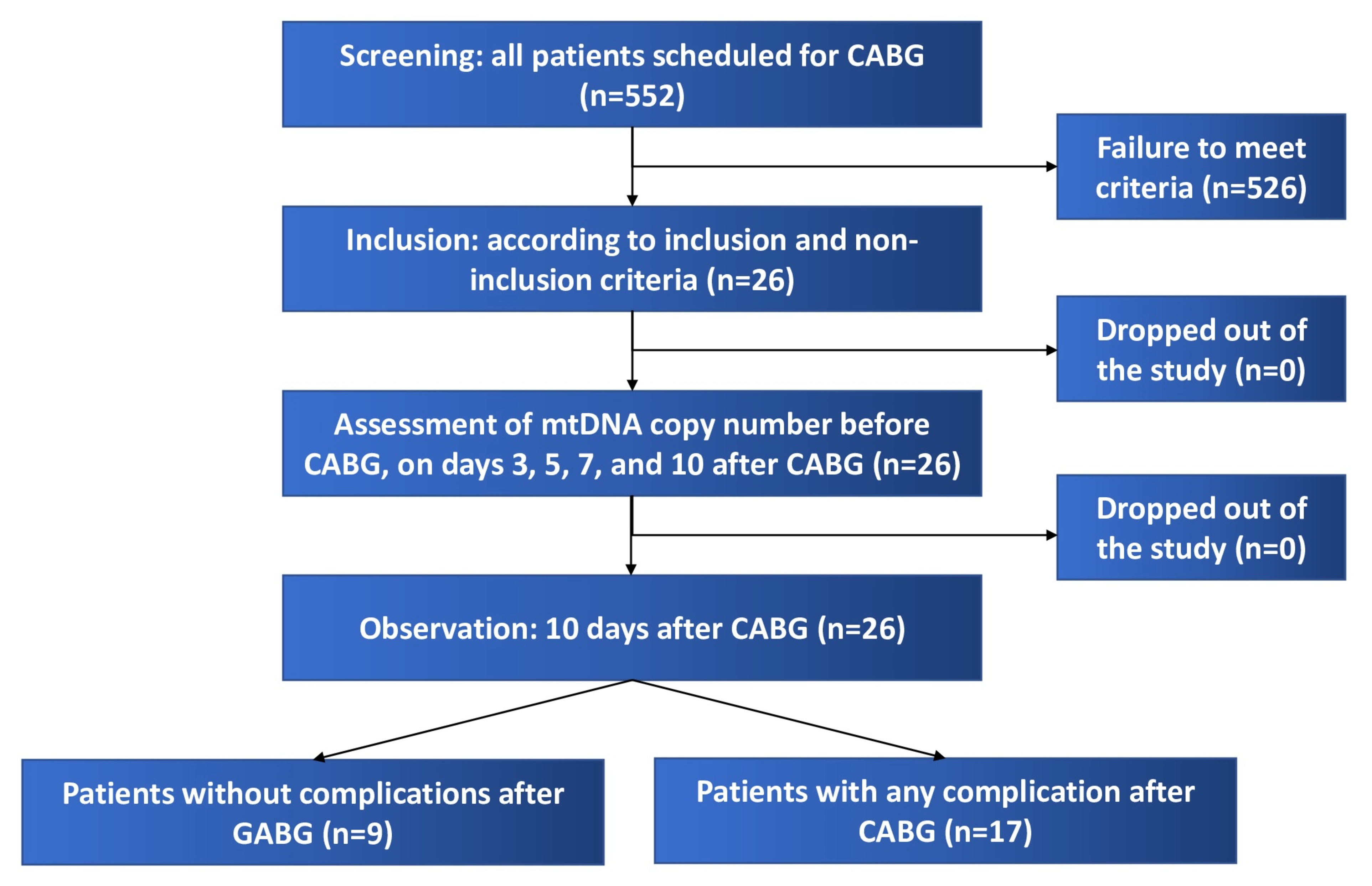 Figure 1. Study design flow diagram.
All patients voluntarily provided written informed consent for the use of blood and plasma samples. The Ethics Committee at the Institute for Cardiology Research of the Tomsk National Medical Research Center approved this study (approval no. 241 of March 9, 2023), which was conducted in accordance with the Declaration of Helsinki (2013). Blood plasma and peripheral blood mononuclear cells (PBMCs) were collected from patients at baseline (before CABG) and on days 1, 3, 5, 7, and 10 postoperatively. The study was conducted using equipment at the Medical Genomics Shared Use Center of the Institute for Cardiology Research, Tomsk, Russia.
DNA preparation
To obtain DNA from PBMCs, whole blood was washed twice with ACK lysis buffer prepared according to the protocol described by AAT Bioquest [14]. DNA was isolated from 300 µL of blood using the D-Blood kit (Biolabmix). The quantity and quality of the isolated DNA samples were assessed using a Thermo Scientific NanoDropTM 8000 spectrophotometer. DNA degradation was assessed by electrophoresis in a 1% agarose gel. DNA samples were diluted to a working concentration of 20 ng/µL using 1X TE buffer with 0.1 mM EDTA (SibEnzyme). For digestion, 20 ng of DNA was treated with 1 unit of Thermo Scientific HinfI restriction enzyme and Thermo Scientific 1X Tango buffer for 1 h at 37 °C. Subsequently, 1 ng of digested DNA per 20 µL of reaction mixture was used for digital polymerase chain reaction (PCR).
Due to the low quality, low reproducibility, and high variability of results obtained when measuring mtDNA in plasma using direct droplet digital PCR (dddPCR) by adding 1 µL of plasma to the reaction mixture, this study focused on the isolation and purification of circulating cell-free DNA (cfDNA).
Plasma was collected using GRADBIOMED® GBM scf-DNA GBM vacuum tubes specifically designed for cfDNA analysis. A MagPure Circulating DNA Kit (Magen Biotech) was utilized to extract cfDNA from 300 µL of plasma according to the manufacturer’s protocol, and cfDNA samples were resuspended in 50 µL of elution buffer. Quantification of cfDNA was performed fluorometrically using the Qubit dsDNA HS Assay Kit (Thermo Scientific) on a Thermo Scientific Qubit 3 fluorometer. Then cfDNA samples were subjected to a 200-fold dilution, and 1 μL of the diluted cfDNA was used for mtDNA quantification by digital PCR (dPCR).
Measuring mt-DNA copy number by digital PCR
This study utilized primers and probes developed and patented by our research group for mtDNA quantification using dPCR. These primers were designed to quantify mtDNA by measuring the copy number of the MT-RNR2 gene, using the ACTB gene as a reference. The patent for these primers was registered under the number RU2755663C1 [15].
The standard 20 µL droplet digital PCR (ddPCR) reaction mixture contained two pairs of primers for MT-RNR2 and ACTB at final concentrations of 500 nM and 250 nM, respectively, and 1 µL of prepared DNA. All manipulations were performed using low-adhesion consumables. 2X Digital PCR Mastermix for Probes (RainSure Scientific) was used as the master mix, and Generation Oil for Probes (RainSure Scientific) was utilized to generate droplets. Further preparation of the ddPCR assay was performed according to the manufacturer’s protocol for the QX200 Droplet Digital PCR System (Bio-Rad).
The amplification program on the compatible CFX96 Touch Real-Time PCR Detection System (Bio-Rad Scientific) was used according to the manufacturer’s recommendations, with modifications, viz., a ramp rate of 1.5 °C/sec and an additional overnight storage step at 4 °C in the refrigerator to stabilize the droplets and increase the yield of read droplets. Results were analyzed using QuantaSoft v.1.7.4 software.
Quantification of mtDNA in PBMCs was performed per cell, i.e., the number of MT-RNR2 copies in 20 µL/the number of ACTB copies in 20 µL×2 (diploid chromosome set). The amount of mtDNA was calculated per 1 mL of blood plasma, as follows: the resulting number of MT-RNR2 copies in 20 µL of the reaction mixture, multiplied by 200 (dilution factor) and by 50 µL (eluate volume), divided by 300 µL of plasma used, and multiplied by 1,000.
Measuring blood level of cytochrome c
The concentration of cytochrome c (ng/mL) in serum was determined by ELISA using a CusaBio kit (USA) on equipment at the Medical Genomics Shared Use Center of the Tomsk National Medical Research Center. This analysis was performed before CABG, as well as 3 and 10 days after CABG.
Definition of postoperative complications
As postoperative complications, we considered the following issues: infectious complications (e.g., surgical site infections, pneumonia, urinary tract infections, and deep sternal wound infections); acute kidney injury (defined as an acute decline in kidney function resulting in an increase in serum creatinine and/or a decrease in urine output [16]); postpericardiotomy syndrome (diagnosed when patients had at least two of the following issues: fever without an alternative cause, pleuritic chest pain, friction rub, new or worsening pleural effusion, and new or worsening pericardial effusion [17]); acute myocardial infarction (diagnosed according to the Fourth Universal Definition of Myocardial Infarction [18]); stroke confirmed by imaging; and new-onset atrial fibrillation.
Statistical analysis
We used IBM SPSS Statistics v.21 for data analysis. The Shapiro-Wilk test was utilized to assess normal distribution. Given the small sample size and the lack of normal distribution for most variables, nonparametric methods were employed for statistical analysis. Continuous variables are presented as medians (Q25-Q75). Categorical data are presented as counts (percentages). The Mann-Whitney U test was applied to intergroup comparisons of continuous variables (independent groups), while the Wilcoxon signed-rank test was used for paired samples. Spearman’s rank correlation coefficient was used to assess correlations. Categorical variables were compared using the χ² test or Fisher’s exact test (for small sample sizes). The median percentage change in mtDNA copy number from the preoperative period to 10 days after CABG was calculated as follows:
 (1)
(1)
The median percentage change in cytochrome c concentration from the preoperative period to 10 days after CABG was calculated as:
 (2)
(2)
The relationship between this change in mtDNA copy number and postoperative complications was assessed using receiver operating characteristic (ROC) curve analysis (area under the curve [AUC]). For multiple comparisons of parameters (changes in parameters on days 3, 5, 7, and 10 after CABG), p<0.01 was considered statistically significant, taking into account the Bonferroni correction. In all other cases, p<0.05 was considered statistically significant.
Results
This study included 26 patients with CAD who underwent CABG. The median age was 67 (58-71) years, and 92.3% of patients were men. Detailed patient demographics are presented in Table S1 of the Supplementary Materials section. Plasma mtDNA copy numbers preoperatively and at 3, 5, 7, and 10 days postoperatively are shown in Table 1 and Figure 2.
Table 1. Plasma mtDNA levels before and after coronary artery bypass grafting
|
Parameter |
mtDNA copy number per 1 mL of plasma (in 106/mL) |
p-value (Wilcoxon signed-rank test) |
|||||
|
Median |
(Q25-Q75) |
Background |
Day 3 |
Day 5 |
Day 7 |
Day 10 |
|
|
Background |
5.12 |
(3.35-7.44) |
- |
0.005 |
0.852 |
0.024 |
0.001 |
|
Day 3 |
2.65 |
(1.88-3.04) |
- |
- |
0.017 |
0.005 |
0.005 |
|
Day 5 |
4.04 |
(2.28-7.00) |
- |
- |
- |
0.005 |
<0.001 |
|
Day 7 |
6.50 |
(3.47-11.31) |
- |
- |
- |
- |
0.067 |
|
Day 10 |
10 |
(7.2-13.4) |
- |
- |
- |
- |
- |
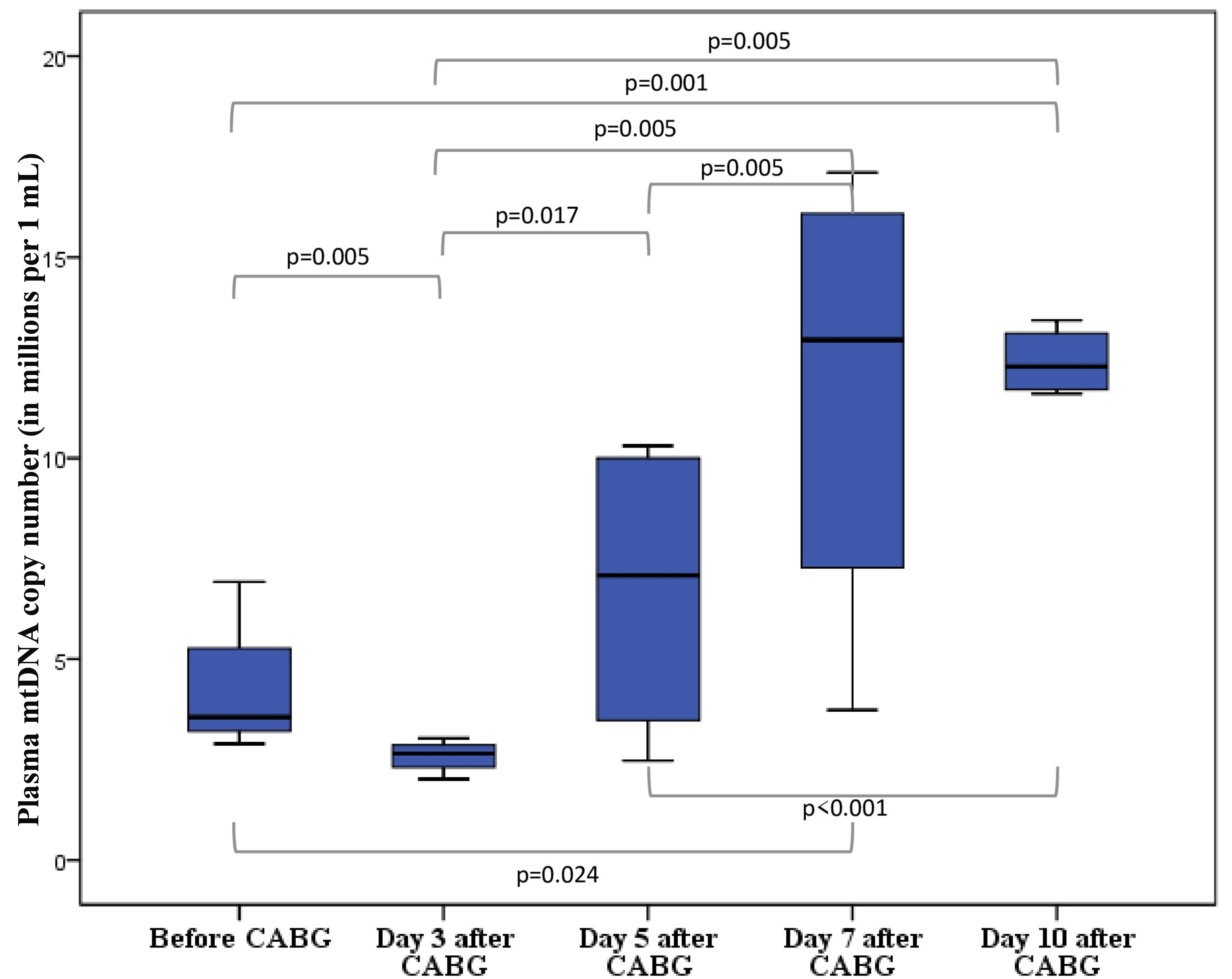
Figure 2. The number of mtDNA copies in blood plasma in patients (n=26) who underwent coronary artery bypass grafting (CABG) using a cardiopulmonary bypass machine and cardioplegia. Blood samples were collected before CABG and at 3, 5, 7, and 10 days after CABG. mtDNA copy number was measured using dPCR and expressed as millions per 1 mL. Data are presented as boxplots showing median values, 25th and 75th percentiles, and minimum and maximum values. P-values for pairwise comparisons using the Wilcoxon signed-rank test are shown below. P-values <0.01 were considered statistically significant.
The results confirmed a postoperative increase in plasma mtDNA copy number: from a median of 2.65 million copies/mL (IQR: 1.88-3.04) at 72 hours postoperatively to a median of 10 million copies/mL (IQR: 7.2-13.4) at 10 days postoperatively (p=0.005). Furthermore, plasma mtDNA copy number was significantly higher at 10 days postoperatively than before CABG (p=0.001). We also quantified the number of mtDNA copies per PBMC (Table 2, Figure 3).
Table 2. Number of mtDNA copies per peripheral blood mononuclear cell (PBMC) before coronary artery bypass grafting and at 3, 5, 7, and 10 days postoperatively
|
Parameter |
mtDNA-copy number per PBMC |
p-value (Wilcoxon signed-rank test) |
|||||
|
Median |
(Q25-Q75) |
Background |
Day 3 |
Day 5 |
Day 7 |
Day 10 |
|
|
Background |
79 |
(63.5-95.2) |
- |
0.689 |
0.689 |
0.867 |
0.346 |
|
Day 3 |
65 |
(58-88) |
- |
- |
0.401 |
0.475 |
0.123 |
|
Day 5 |
81 |
(59.5-107.5) |
- |
- |
- |
0.556 |
0.198 |
|
Day 7 |
82 |
(67-94) |
- |
- |
- |
- |
0.411 |
|
Day 10 |
86 |
(70-114) |
- |
- |
- |
- |
- |
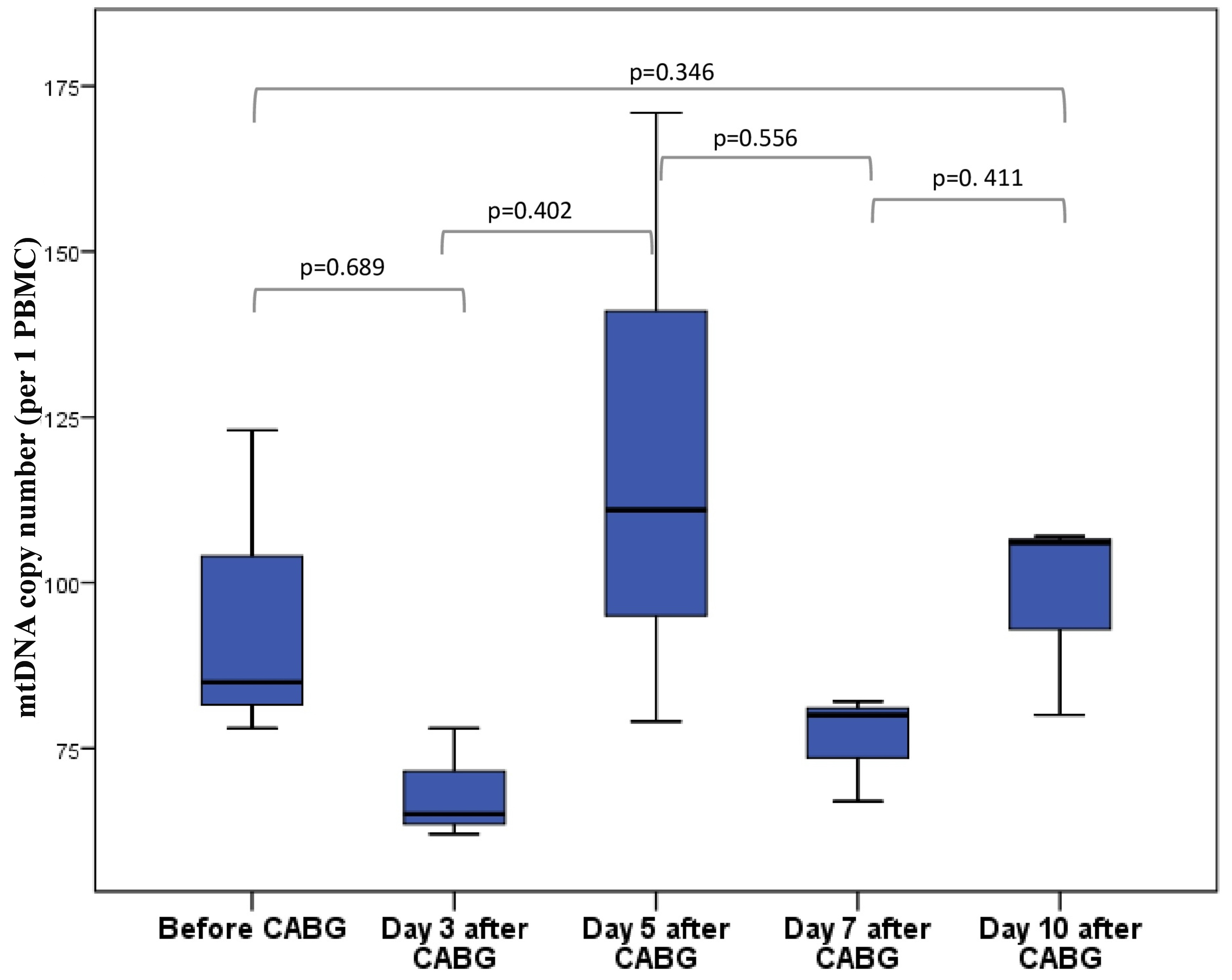
Figure 3. The number of mtDNA copies per peripheral blood mononuclear cell (PBMC) in patients (n=26) who underwent coronary artery bypass grafting (CABG) using a cardiopulmonary bypass machine and cardioplegia. PBMC specimens were collected before CABG and at 3, 5, 7, and 10 days after CABG. mtDNA copy number was measured using dPCR and expressed as millions per 1 PBMC. Data are presented as boxplots showing median values, 25th and 75th percentiles, and minimum and maximum values. P-values for pairwise comparisons using the Wilcoxon signed-rank test are shown below. P-values <0.01 were considered statistically significant.
The mtDNA copy number in PBMCs remained stable throughout the perioperative period, without statistically significant changes (p>0.05). We also assessed the correlation between the mtDNA copy number in PBMCs and several clinical parameters. No significant correlations were observed with body mass index (r=0.053, p=0.800), waist circumference (r=0.370, p=0.130), or blood glucose levels (r=-0.261, p=0.199).
The median percentage change in mtDNA copy number, calculated using formula (1), was 116% (IQR: 9.6-267%). This parameter did not correlate with patient age (r=-0.166, p=0.449), NT-proBNP concentration (r=0.162, p=0.521), left ventricular ejection fraction (LVEF) (r=0.233, p=0.296), cardiopulmonary bypass time (r=0.027, p=0.907), or aortic cross-clamping time (r=0.408, p=0.159). Furthermore, mtDNA copy number did not differ significantly between men and women (p=0.743).
Given the increased mtDNA copy number in plasma alongside the stable mtDNA copy number in PBMCs, we hypothesized that the increased mtDNA in plasma is associated with further cellular injury in the postoperative period. Therefore, we analyzed the relationship between this parameter and postoperative complications during this hospitalization. Indeed, patients with any complications (infection, acute kidney injury, postpericardiotomy syndrome, acute myocardial infarction, stroke, atrial fibrillation) (n=17) had a significantly higher median change in plasma mtDNA copy number (198%; IQR: 101; 321%) vs. patients without complications (n=9) (11%; IQR: -36; 127%; p=0.008) (Figure 4, Table 3). However, no significant differences in mtDNA copy number changes were found for individual categories of complications: infection (p=0.520), acute kidney injury (p=0.129), postpericardiotomy syndrome (p=0.806), acute myocardial infarction (p=0.583), stroke (p=0.392), or atrial fibrillation (p=0.463). In addition, we identified a group of patients with serious complications such as deep sternal wound infection, stroke, or myocardial infarction (n= 3). The plasma mtDNA copy number change in this group ranged from -44 to 453%. We did not obtain statistically significant results in this group due to the large spread of data and the small number of patients with serious complications (p=0.263). However, statistically significant differences in mtDNA copy number change remained between patients with less severe complications and those without complications (p=0.009). The incidence of postoperative complications is presented in Table 2 of the Supplementary Materials section. A comparison of clinical data for patients with and without complications after CABG using a cardiopulmonary bypass machine is presented in Table S3 (Supplementary Materials). The groups did not differ statistically significantly in the main clinical parameters.
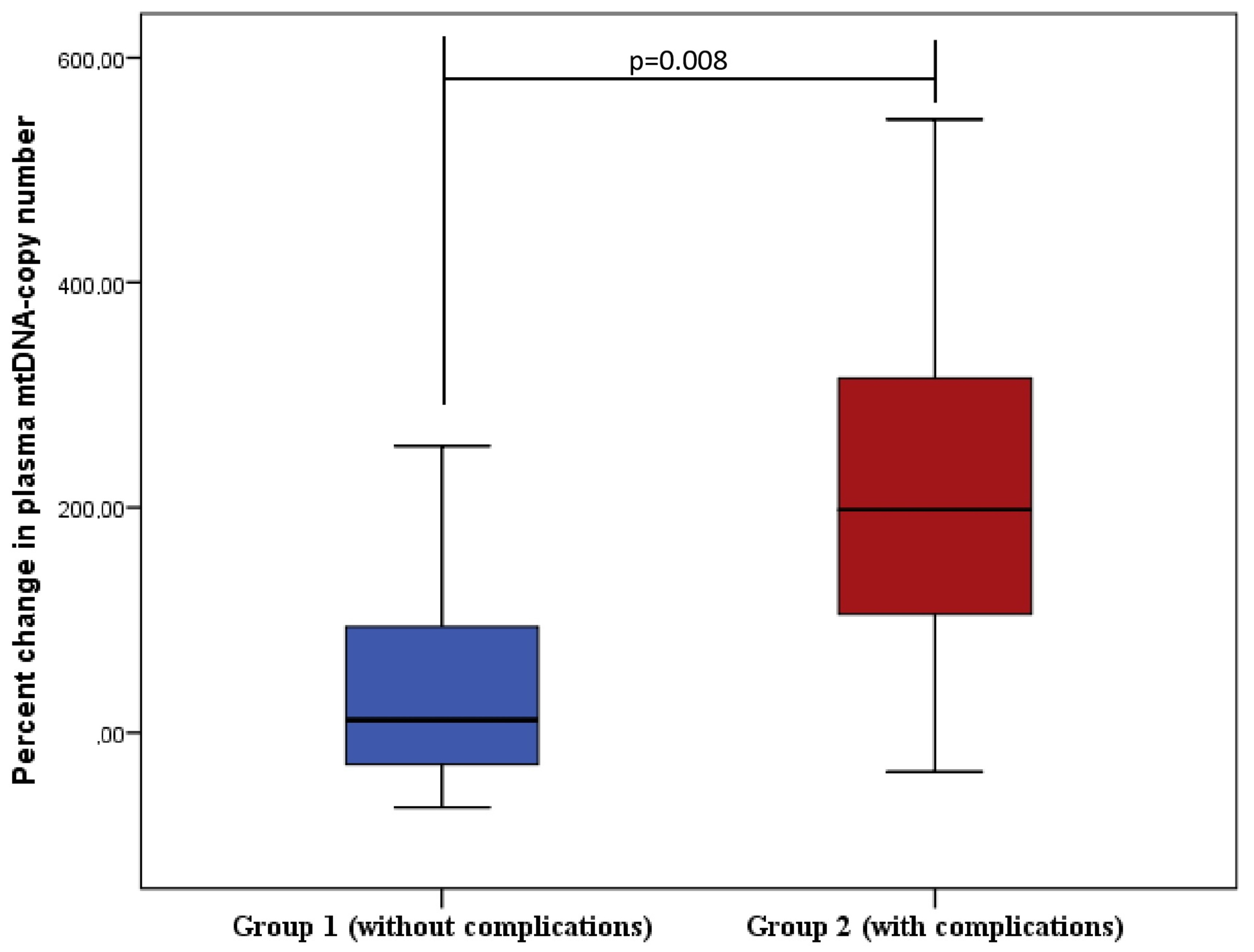
Figure 4. Change in plasma mtDNA copy number from preoperative value to Day 10 days after coronary artery bypass grafting (CABG) in patients with and without postoperative complications. Group 1 includes patients without any complications (n=9), while Group 2 encompasses patients with complications (n=17), including infection, acute kidney injury, postpericardiotomy syndrome, acute myocardial infarction, stroke, and atrial fibrillation. The p-value for comparison using the Wilcoxon signed-rank test for paired samples is 0.008. Data are presented as boxplots showing median values, 25th and 75th percentiles, and minimum and maximum values; 95% confidence interval for the mean change in mtDNA copy number: 95% CI [-28; 125] in Group 1, 95% CI [130; 298] in Group 2.
Table 3. Changes in mtDNA copy number in patients with and without complications after coronary artery bypass grafting (CABG)
|
Parameter |
Without complications (n=9) |
With complications (n=17) |
р |
|
mtDNA copy number per 1 mL of blood plasma before CABG, (106/mL) |
5.05 (3.2-7.9) |
5.2 (3.1-6.7) |
0.718 |
|
mtDNA copy number per 1 mL of blood plasma on Day 10 after CABG, (106/mL) |
7.25 (6-11.4) |
11.6 (8-14.6) |
0.147 |
|
mtDNA copy number change, % |
11 ((-36)-127) |
198 (101-321) |
0.008 |
The median plasma mtDNA copy number did not differ statistically significantly between patients with complications (5.05 million copies/mL; IQR: 3.2-7.9) and without complications (5.2 million copies/mL; IQR: 3.1-6.7; p=0.718). ROC curve analysis showed that an increase in plasma mtDNA copy number of 30% or more vs. the preoperative value was associated with the development of postoperative complications, with a sensitivity of 0.88 and a specificity of 0.67 (Figure 5).
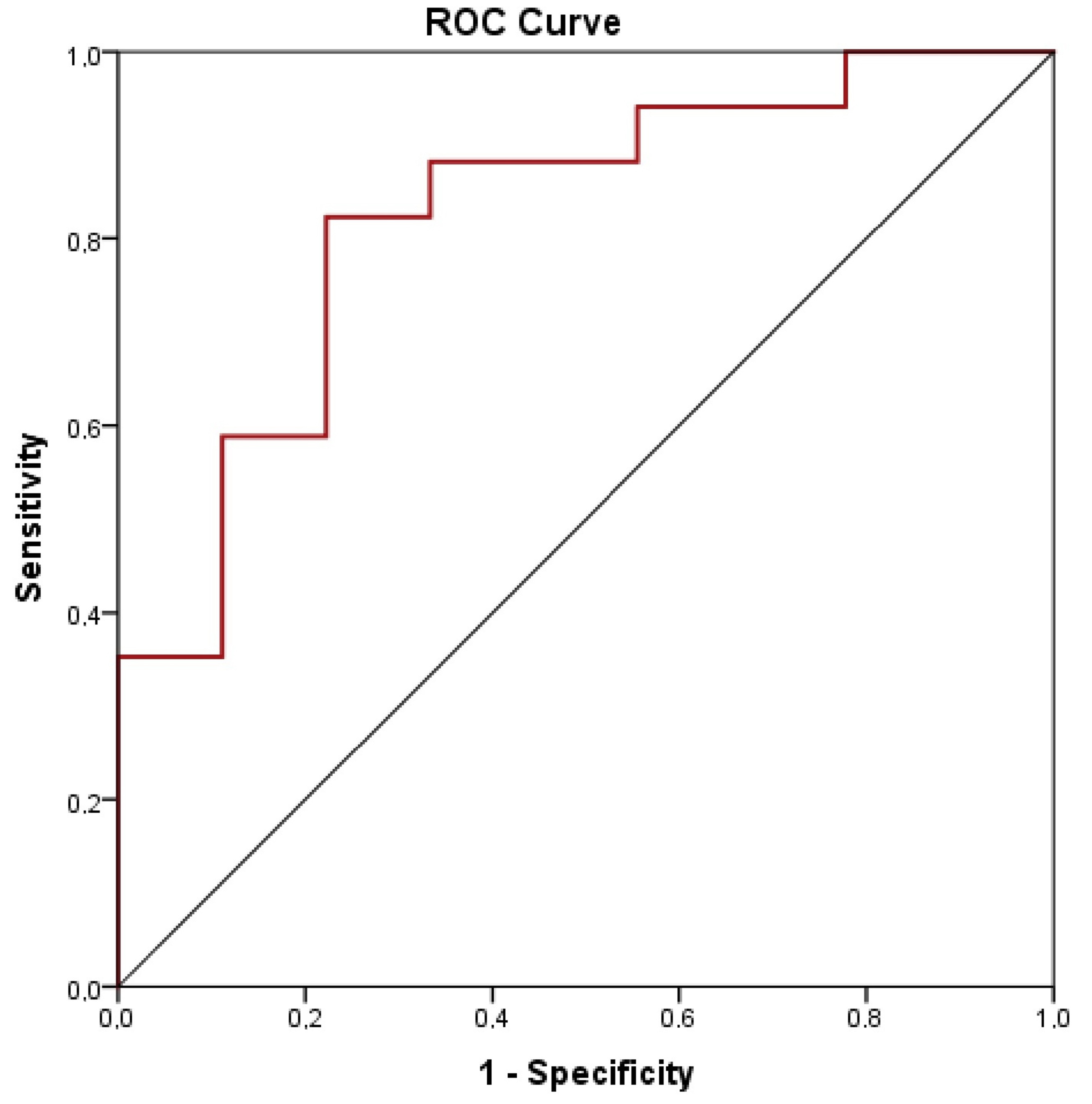
Figure 5. Receiver operating characteristic (ROC) curve for predicting postoperative complications based on changes in plasma mtDNA copy number. The curve was constructed using data from patients who experienced postoperative complications (including infection, acute kidney injury, postpericardiotomy syndrome, acute myocardial infarction, stroke, and atrial fibrillation). A 30% increase in plasma mtDNA copy number was associated with postoperative complications, with a sensitivity of 0.88, a specificity of 0.67, and AUC of 0.824 (95% CI: 0.653–0.994; p=0.008).
To confirm that patients with postoperative complications experience greater mitochondrial destruction, we analyzed cytochrome c concentrations before CABG and on days 3 and 10 after CABG (Table 4).
Table 4. Changes in cytochrome с concentration in patients with and without complications after coronary artery bypass grafting (CABG)
|
Parameter |
Without complications (n=9) |
With complications (n=17) |
р |
|
Cytochrome c levels before CABG, ng/mL |
25.1 (18.2; 32.6) |
24.8 (14; 31.7) |
0.779 |
|
Cytochrome c levels on Day 3 after CABG, ng/mL |
32.4 (25.8; 39) |
32.5 (22.3; 49) |
0.968 |
|
Cytochrome c levels on Day 10 after CABG, ng/mL |
27.4 (18.8; 32.1) |
34.2 (25.6; 39.3) |
0.282 |
|
Cytochrome c level change, % |
4.1 (-34; 54) |
31.7 (6.3; 92.8) |
0.039 |
Thus, cytochrome c levels before CABG did not differ between the groups with and without complications. By postoperative Day 3, we recorded an increase in cytochrome c concentrations in both groups (p=0.968 when comparing Group 1 with Group 2). Subsequently, cytochrome c concentration remained high in the group with complications, while decreasing in the group without complications 10 days after CABG. We calculated the change in cytochrome c level from the preoperative period to postoperative Day 10. In the group without complications, the change in cytochrome c concentration was 4.1% (-34; 54), while in the group with complications it was 31.7% (6.3; 92.8); the differences were statistically significant (p=0.039).
Discussion
This study is the first to demonstrate the dynamics of plasma mtDNA copy numbers over 10 days after on-pump CABG. We observed a slight decrease in plasma mtDNA copy numbers vs. preoperative levels on postoperative Day 3, which is consistent with published data [8]. Subsequently, a statistically significant increase in plasma mtDNA copy numbers was observed for 10 days after surgery.
According to the published sources, an increase in plasma mtDNA copy numbers during the perioperative period may be associated with mechanical cellular trauma, the use of cardiopulmonary bypass in cardiac surgery, and reperfusion. However, after surgery, mtDNA copy numbers should return to normal within 24-72 hours [7, 8, 19].
Due to the design of our study, we were able to observe an increase in plasma mtDNA copy numbers 10 days after cardiac surgery under cardiopulmonary bypass. Given that mechanical cell damage and the use of pump-activated circulation do explain this change, we hypothesize that ongoing cell damage caused by inflammation after cardiac surgery leads to the release of mtDNA, which in turn enhances inflammatory processes [6]. By now, mtDNA has been extensively studied as a danger signal. It is a molecule that alerts the body by activating an inflammatory program in target cells [12]; mtDNA released from the mitochondria is a potent activator of cyclic GMP-AMP synthase, which leads to stimulation of the subsequent synthesis of cytokines such as interferon-β1, IL-6, and tumor necrosis factor [10].
Progressive inflammation, in turn, can trigger the development and progression of complications after on-pump CABG, primarily atrial fibrillation and postpericardiotomy syndrome, which significantly worsen the postoperative course and prolong hospitalization [20, 21].
We discovered that the greatest increase in plasma mtDNA copy number occurred in patients with postoperative complications, including infections, acute kidney injury, postpericardiotomy syndrome, acute myocardial infarction, stroke, and atrial fibrillation. Plasma mtDNA copy number 10 days after surgery was not statistically significantly associated with postoperative complications. These results are consistent with previous published studies showing that a more than twofold increase in plasma mtDNA copy number within two days after CABG is associated with a higher risk of complications such as atrial fibrillation and infections [6]. This may be due to the adverse effects of mtDNA DAMPs [22].
Other studies have reported an association between preoperative mtDNA copy number in the blood and the development of atrial fibrillation [6, 9]. We did not observe this pattern in our cohort, possibly due to the inclusion of patients with severe HF, which could have resulted in elevated preoperative plasma mtDNA copy number. Another possible explanation is the small number of patients who developed atrial fibrillation (n=6), which may have limited statistical power.
We demonstrated that an increase in plasma mtDNA copy number of 30% or more is associated with the development of postoperative complications with a sensitivity of 88% and a specificity of 67% (ROC analysis). This indirectly confirms the presence of a pathological association between them.
To confirm this association, we examined an additional marker, cytochrome c, which enters the bloodstream when mitochondria are destroyed. Indeed, changes in cytochrome c concentrations 10 days after CABG were associated with the development of postoperative complications. Previous studies reported that cytochrome c levels increase in patients after cardiac arrest, and to a greater extent in those patients who died, which proves its association with cardiomyocyte damage [23]. The association of cytochrome c level with mitochondrial damage was also demonstrated in experimental studies [13]. Another study [24] demonstrated no changes in cytochrome c concentrations 6 hours after CABG. However, we could not find any published sources examining changes in cytochrome c concentration 10 days after CABG. In our group of patients with complications after CABG, an increase in cytochrome c levels was observed by Day 3 after surgery and remained high until Day 10. In the group of patients without complications, a decrease in cytochrome c concentration was recorded from Day 3 to Day 10. These data, along with an increase in plasma mtDNA copy number, indicate ongoing cellular and mitochondrial damage in the postoperative period in patients with complications.
The complication rate in our study was 65.4%, which is quite high, compared with published data, including more severe cohort of patients in the STICH study [25], where the complication rate was 31%. However, the STICH study did not take into account the development of postpericardiotomy syndrome, which was one of the most common complications in our group. Furthermore, if we consider solely serious complications such as deep chest infection, stroke, and myocardial infarction, the incidence of such complications was low in our group (11.5%). The remaining complication cases were associated with less serious events. We believe that the development of complications in our study was not related to the specific surgical procedure, as the main characteristics of cardiac surgery did not differ between patients with and without complications.
We attribute the high complication rate to the baseline severity of the clinical condition in our patients, who had a reduced left ventricular ejection fraction, and over 80% of whom had a history of myocardial infarction. One in four patients experienced HF decompensation within a year prior to surgery.
Many studies explored the correlation between mtDNA copy number abnormalities and cardiovascular disease. However, their results are contradictory. Studies analyzing the relationship between mtDNA copy number and complications after cardiac surgery [6-8] were limited to a 72-hour period when changes in mtDNA copy number could be caused by mechanical trauma, which could affect the measured parameter. Some of these studies also had small sample sizes. Although other studies included large numbers of patients, they only examined preoperative values [9], which is insufficient to show how this parameter changes after on-pump CABG. Our advantage is that we studied mtDNA copy number in a series and were able to assess changes in this parameter for 10 days after on-pump CABG. Therefore, our data are novel and may inspire larger studies in this area.
Thus, our results indicate an increase in plasma mtDNA copy number unrelated to mechanical trauma or cardiopulmonary bypass. This suggests that reducing plasma mtDNA copy number may reduce inflammation and the risk of complications. Mitochondria are increasingly considered as a target for new cardiovascular drug therapies [19], and our study adds new evidence to this concept.
In our study, the mtDNA copy number in PBMCs did not change either before surgery or on days 3, 5, 7, and 10 after CABG. According to the literature, a reduction in the mtDNA copy number in PBMCs is associated with metabolic disorders [26]; however, in our study, no statistically significant correlations were found between this parameter and glycemia levels, body mass index, or waist circumference. Low mtDNA copy number in PBMCs in the study cohort may indicate a high baseline cardiovascular risk in patients with HF and CAD [27-29]. Furthermore, the dynamics of mtDNA copy number in PBMCs was not previously studied in patients after CABG. Hence, we showed for the first time that the number of mtDNA copies in the PBMC does not change statistically significantly after CABG, which may indirectly indicate a decrease in the reparative activity of mtDNA in PBMCs in our group of patients [30].
Limitations
The main limitation of our study was its small sample size. However, the number of patients included was sufficient to demonstrate the dynamics of mtDNA copy number in peripheral blood and identify statistically significant associations between this biomarker and the development of postoperative complications. It is worth noting that most complications in our patients were minor, while deep sternal wound infections, strokes, and myocardial infarctions were recorded in just three patients. As a result, we cannot distinguish between minor and major complications. Combining all complications into one group for analysis is a significant limitation, as different complications have different etiologies. This could be the subject of further research.
Another issue related to the small number of patients is that 90% of them were men. This fact should be taken into account when extrapolating the results to the general population undergoing on-pump CABG.
One more limitation of our study was the inability to calculate the sample size before its onset, since, to our knowledge, this is the first study to analyze the dynamics of mtDNA copy number over 10 days after coronary artery on-pump CABG and to examine the association between changes in mtDNA copy number and postoperative complications.
Conclusion
Plasma mtDNA copy number decreases to preoperative levels on Day 3 after CABG. Subsequently, the mtDNA copy number in peripheral blood plasma increases by Day 10 after surgery. Based on the results of our pilot study, an increase in plasma mtDNA copy number of 30% or more 10 days after CABG vs. preoperative levels is associated with the development of postoperative complications, which are primarily less serious. The number of mtDNA copies in PBMCs does not change after CABG using a cardiopulmonary bypass machine.
Author contributions
GAA and KEA designed the study and analyzed the data. SAA performed the study of mtDNA copy number. KEA and TOV prepared the manuscript. KMU and SEE provided assistance and advice on data collection and analysis. BYI and BAE performed coronary angiography and participated in data analysis. All authors made editorial revisions to the manuscript. All authors read and approved the final version of the manuscript.
Clinical trial registration
The study protocol was registered on ClinicalTrials.gov: NCT05770349. Date of registration: February 21, 2023.
Funding
The study was supported by grant No. 23-75-00009 from the Russian Science Foundation, https://rscf.ru/project/23-75-00009/
Ethical statement
The study protocol was approved by the Ethics Committee at the Institute for Cardiology Research of the Tomsk National Medical Research Center (Protocol No. 241 of March 9, 2023).
Informed consent statement
Informed consent was obtained from all study participants.
Data availability
The datasets used in this study are available from the corresponding author upon reasonable request.
AI use statement
We did not use any AI tools or technologies in the preparation of this manuscript.
Supplementary Materials
Table S1. Baseline demographic and clinical characteristics of patients before CABG
|
Parametrs |
n=26 |
|
Age, years |
67 (58, 71) |
|
Gender: male, n (%) |
24 (92.3) |
|
Angina, n(%) |
26 (100) |
|
Acute decompensated heart failure in the last 12 months, n (%) |
6 (23.1) |
|
Shortness of breath, n (%) |
26 (100) |
|
Edema, n (%) |
2 (7.7) |
|
Liver enlargement, n (%) |
1 (3.8) |
|
Moist rales, n (%) |
2 (7.7) |
|
History of myocardial infarction, n (%) |
21 (80.8) |
|
NYHA class heart failure |
2 (2, 3) |
|
Hypertension, n(%) |
26 (100) |
|
Stroke, n (%) |
2 (7.7) |
|
Atrial fibrillation, n (%) |
5 (19.2) |
|
Current smoker, n (%) |
12 (46.2) |
|
Obesity, n (%) |
12 (46.2) |
|
Systolic blood pressure, mm Hg |
130 (130, 140) |
|
Diastolic blood pressure, mm Hg |
80 (74, 82) |
|
Heart rate, b.p.m |
68 (61, 75) |
|
Left ventricular ejection fraction, % |
44 (32, 46) |
|
NTproBNP, pg/ml |
142 (74, 298) |
|
Renin-angiotensin-aldosterone inhibitors, % |
25 (96.2) |
|
Beta blockers, % |
22 (84.6) |
|
Mineralocorticoid receptor antagonists, % |
17 (65.4) |
|
Sodium glucose cotransporter type 2 inhibitors, n (%) |
18 (69.2) |
|
Diuretics, n (%) |
20 (77) |
|
Statins, n (%) |
26 (100) |
Table S2. Complications after CABG surgery in the study cohort
|
Parametrs |
n=26 |
|
Infectious complications, n (%) |
10 (38.5) |
|
Acute kidney injury, n (%) |
1 (3.8) |
|
Post-pericardiotomy syndrome, n (%) |
8 (30.7) |
|
Myocardial infarction, n (%) |
1(3.8) |
|
Imaging-confirmed stroke, n (%) |
1 (3.8) |
|
Newly diagnosed atrial fibrillation, n (%) |
6 (23.1) |
|
Any complications |
17 (65.4) |
Table S3. Baseline demographic and clinical characteristics of patients with and without complications
|
Parametrs |
Without complications (n=9) |
With complications (n=17) |
р |
|
Age, years |
66 (51.5; 70) |
68 (58; 71) |
0.426 |
|
Gender: male; n (%) |
9 (100) |
15 (88.2) |
0.529 |
|
Angina; n(%) |
9 (100) |
17 (100) |
1.0 |
|
Acute decompensated heart failure in the last 12 months, n (%) |
2 (22.2) |
4 (23.5) |
0.940 |
|
Shortness of breath, n (%) |
9 (100) |
17 (100) |
1.0 |
|
Edema; n (%) |
0 |
2 (11.8) |
0.285 |
|
Liver enlargement; n (%) |
0 |
1 (5.9) |
0.459 |
|
Moist rales; n (%) |
0 |
2 (11.8) |
0.285 |
|
History of myocardial infarction; n (%) |
7 (77.8) |
14 (82.4) |
0.778 |
|
NYHA class heart failure |
2 (2; 3) |
2 (2; 3) |
0.934 |
|
Hypertension ; n(%) |
9 (100) |
17 (100) |
1.0 |
|
Stroke; n (%) |
0 |
2 (11.8) |
0.285 |
|
Atrial fibrillation; n (%) |
2 (22.2) |
3 (17.7) |
0.779 |
|
Current smoker; n (%) |
6 (66.7) |
6 (35.3) |
0.369 |
|
Obesity; n (%) |
4 (44.4) |
8 (47) |
0.790 |
|
Systolic blood pressure, mm Hg |
130 (130; 140) |
130 (120; 140) |
0.522 |
|
Diastolic blood pressure, mm Hg |
80 (78; 82) |
80 (80; 84) |
0.678 |
|
Heart rate, b.p.m |
68 (61; 81.5) |
67.5 (60.5; 75) |
0.677 |
|
Left ventricular ejection fraction, % |
39 (30; 49) |
44.5 (34; 49.5) |
0.846 |
|
NTproBNP, pg/ml |
129 (36.5; 223) |
155 (75; 316) |
0.445 |
|
Renin-angiotensin-aldosterone inhibitors, % |
8 (88.9) |
17 (100) |
0.162 |
|
Beta blockers, % |
8 (88.9) |
14 (82.4) |
0.661 |
|
Mineralocorticoid receptor antagonists, % |
6 (66.7) |
11 (64.7) |
0.921 |
|
Sodium glucose cotransporter type 2 inhibitors, n (%) |
6 (66.7) |
12 (70.6) |
0.837) |
|
Diuretics, n (%) |
6 (66.7) |
14 (87.5) |
0.367 |
|
Statins, n (%) |
9 (100) |
17 (100) |
1.0 |
|
On-pump time, min |
92 (89-104.5) |
97.5 (82.5-112.25) |
0,755 |
|
Aortic cross-clamp time, min |
56 (43.5; 66) |
47.5 (41; 59.25) |
0,243 |
|
Number of bypass grafts, n (%) 2 3 4 |
3 (33.3) 5 (55.6) 1 (11.1) |
4 (23.5) 10 (58.8) 3 (17.7) |
0,733
|
- Atici AE, Crother TR, Noval Rivas M. Mitochondrial quality control in health and cardiovascular diseases. Front Cell Dev Biol 2023; 11: 1290046. https://doi.org/10.3389/fcell.2023.1290046.
- Guo Z, Tian Y, Liu N, Chen Y, Chen X, Yuan G, et al. Mitochondrial stress as a central player in the pathogenesis of hypoxia-related myocardial dysfunction: New insights. Int J Med Sci 2024; 21(13): 2502-2509. https://doi.org/10.7150/ijms.99359.
- Alves-Figueiredo H, Silva-Platas C, Lozano O, Vázquez-Garza E, Guerrero-Beltrán CE, Zarain-Herzberg A, et al. A systematic review of post-translational modifications in the mitochondrial permeability transition pore complex associated with cardiac diseases. Biochim Biophys Acta Mol Basis Dis 2021; 1867(1): 165992. https://doi.org/10.1016/j.bbadis.2020.165992.
- Paraskevaidis I, Kourek C, Farmakis D, Tsougos E. Mitochondrial dysfunction in cardiac disease: The fort fell. Biomolecules 2024; 14(12): 1534. https://doi.org/10.3390/biom14121534.
- Anderson S, Bankier AT, Barrell BG, de Bruijn MH, Coulson AR, Drouin J, et al. Sequence and organization of the human mitochondrial genome. Nature 1981; 290(5806): 457-465. https://doi.org/10.1038/290457a0.
- Sandler N, Kaczmarek E, Itagaki K, Zheng Y, Otterbein L, Khabbaz K, et al. Mitochondrial DAMPs are released during cardiopulmonary bypass surgery and are associated with postoperative atrial fibrillation. Heart Lung Circ 2018; 27(1): 122-129. https://doi.org/10.1016/j.hlc.2017.02.014.
- Baysa A, Fedorov A, Kondratov K, Ruusalepp A, Minasian S, Galagudza M, et al. Release of mitochondrial and nuclear DNA during on-pump heart surgery: Kinetics and relation to extracellular vesicles. J Cardiovasc Transl Res 2019; 12(3): 184-192. https://doi.org/10.1007/s12265-018-9848-3.
- Qin C, Gu J, Hu J, Qian H, Fei X, Li Y, et al. Platelets activation is associated with elevated plasma mitochondrial DNA during cardiopulmonary bypass. J Cardiothorac Surg 2016; 11(1): 90. https://doi.org/10.1186/s13019-016-0481-4.
- Zhang J, Xu S, Xu Y, Liu Y, Li Z, Zhang Y, et al. Relation of mitochondrial DNA copy number in peripheral blood to postoperative atrial fibrillation after isolated off-pump coronary artery bypass grafting. Am J Cardiol 2017; 119(3): 473-477. https://doi.org/10.1016/j.amjcard.2016.10.017.
- Marchi S, Guilbaud E, Tait SWG, Yamazaki T, Galluzzi L. Mitochondrial control of inflammation. Nat Rev Immunol 2023; 23(3): 159-173. https://doi.org/10.1038/s41577-022-00760-x.
- Rucci C, de Simone G, Salathia S, Casadidio C, Censi R, Bordoni L. Exploring mitochondrial DNA copy number in circulating cell-free DNA and extracellular vesicles across cardiovascular health status: A prospective case-control pilot study. FASEB J 2024; 38(10): e23672. https://doi.org/10.1096/fj.202400463R.
- De Gaetano A, Solodka K, Zanini G, Selleri V, Mattioli AV, Nasi M, et al. Molecular mechanisms of mtDNA-mediated inflammation. Cells 2021; 10(11): 2898. https://doi.org/10.3390/cells10112898.
- Radhakrishnan J, Wang S, Ayoub IM, Kolarova JD, Levine RF, Gazmuri RJ. Circulating levels of cytochrome c after resuscitation from cardiac arrest: a marker of mitochondrial injury and predictor of survival. Am J Physiol Heart Circ Physiol 2007; 292(2): H767-75. https://doi.org/10.1152/ajpheart.00468.2006.
- "Quest Calculate™ ACK Lysis Buffer Preparation and Recipe". AAT Bioquest 2026. https://www.aatbio.com/resources/buffer-preparations-and-recipes/ack-lysis-buffer.
- Sleptsov AA, Golubenko MV, Salakhov RR, Nazarenko MS. Method for measuring number of copies of human mitochondrial DNA by digital polymerase chain reaction (Patent No. RU2755663C1). Date of registration: 04/07/2021. Date of publication: 09/20/2021. https://www.elibrary.ru/gwthvi.
- Kidney Disease: Improving Global Outcomes (KDIGO) CKD Work Group. KDIGO 2024 Clinical Practice Guideline for the Evaluation and Management of Chronic Kidney Disease. Kidney Int 2024; 105(4S): S117-S314. https://doi.org/10.1016/j.kint.2023.10.018.
- Imazio M, Brucato A, Ferrazzi P, Spodick DH, Adler Y. Postpericardiotomy syndrome: A proposal for diagnostic criteria. J Cardiovasc Med (Hagerstown) 2013; 14(5): 351-353. https://doi.org/10.2459/JCM.0b013e328353807d.
- Fourth universal definition of myocardial infarction (2018). Rev Esp Cardiol (Engl Ed). 2019; 72(1): 72. English, Spanish. https://doi.org/10.1016/j.rec.2018.11.011.
- Paillard M, Abdellatif M, Andreadou I, Bär C, Bertrand L, Brundel BJJM, et al. Mitochondrial targets in ischaemic heart disease and heart failure, and their potential for a more efficient clinical translation. A scientific statement of the ESC Working Group on Cellular Biology of the Heart and the ESC Working Group on Myocardial Function. Eur J Heart Fail 2025; 27(9): 1720-1736. https://doi.org/10.1002/ejhf.3674.
- Pan T, Jiang CY, Zhang H, Han XK, Zhang HT, Jiang XY, et al. The low-dose colchicine in patients after non-CABG cardiac surgery: A randomized controlled trial. Crit Care 2023; 27(1): 49. https://doi.org/10.1186/s13054-023-04341-9.
- Diakova ML, Shipulin VM, Svirko YS, Gusakova AM, Podoksenov YK, Kamenshchikov NO, et al. Systemic inflammatory response in cardiac surgery: Possibilities of using colchicine. Kardiologiia 2023; 63(7): 39-46. Russian. https://doi.org/10.18087/cardio.2023.7.n2229.
- Paunel-Görgülü A, Wacker M, El Aita M, Hassan S, Schlachtenberger G, Deppe A, et al. fDNA correlates with endothelial damage after cardiac surgery with prolonged cardiopulmonary bypass and amplifies NETosis in an intracellular TLR9-independent manner. Sci Rep 2017; 7(1): 17421. https://doi.org/10.1038/s41598-017-17561-1.
- Cocchi MN, Andersen LW, Rittenberger J, Abella B, Gaieski D, Peberdy MA, et al. Abstract 19510: Cytochrome c levels in post-cardiac arrest patients. Circulation 2015; 132(S3): 19510. https://doi.org/10.1161/circ.132.suppl_3.19510.
- Andersen LW, Liu X, Montissol S, Holmberg MJ, Fabian-Jessing BK, Donnino MW; Center for Resuscitation Science Research Group. Cytochrome c in patients undergoing coronary artery bypass grafting: A post hoc analysis of a randomized trial. J Crit Care 2017; 42: 248-254. https://doi.org/10.1016/j.jcrc.2017.08.006.
- Wrobel K, Stevens SR, Jones RH, Selzman CH, Lamy A, Beaver TM, et al. Influence of baseline characteristics, operative conduct, and postoperative course on 30-day outcomes of coronary artery bypass grafting among patients with left ventricular dysfunction: Results from the surgical treatment for ischemic heart failure (STICH) trial. Circulation 2015; 132(8): 720-730. https://doi.org/10.1161/CIRCULATIONAHA.114.014932.
- Agius R, Pace NP, Fava S. Reduced leukocyte mitochondrial copy number in metabolic syndrome and metabolically healthy obesity. Front Endocrinol (Lausanne) 2022; 13: 886957. https://doi.org/10.3389/fendo.2022.886957.
- Ashar FN, Zhang Y, Longchamps RJ, Lane J, Moes A, Grove ML, et al. Association of mitochondrial DNA copy number with cardiovascular disease. JAMA Cardiol 2017; 2(11): 1247-1255. https://doi.org/10.1001/jamacardio.2017.3683.
- Koller A, Fazzini F, Lamina C, Rantner B, Kollerits B, Stadler M, et al. Mitochondrial DNA copy number is associated with all-cause mortality and cardiovascular events in patients with peripheral arterial disease. J Intern Med 2020; 287(5): 569-579. https://doi.org/10.1111/joim.13027.
- Li X, Liu X, Chen X, Wang Y, Wu S, Li F, et al. Leukocyte mitochondrial DNA copy number and cardiovascular disease: A systematic review and meta-analysis of cohort studies. iScience 2024; 27(9): 110522. https://doi.org/10.1016/j.isci.2024.110522.
- Chan SW, Chevalier S, Aprikian A, Chen JZ. Simultaneous quantification of mitochondrial DNA damage and copy number in circulating blood: A sensitive approach to systemic oxidative stress. Biomed Res Int 2013; 2013: 157547. https://doi.org/10.1155/2013/157547.
Received 7 April 2026, Revised 29 May 2026, Accepted 6 June 2026
© 2026, Russian Open Medical Journal
Correspondence to Elena A. Kuzheleva. E-mail: kea@cardio-tomsk.ru.