

Introduction
Opisthorchiasis is a foodborne parasitic disease caused by the trematode Siberian liver fluke (Opisthorchis felineus Rivolta, 1884), which infects the bile ducts of the liver and pancreatic ducts for up to 30-40 years. The disease is prevalent among residents of Western Siberia and the Urals, and cases have also been reported in other regions as a result of imported infections. An assessment of the risk of O. felineus infection using a multivariate Bayesian geostatistical approach with high spatiotemporal resolution revealed a poverty-related prevalence trend of 46.61% in 2019, corresponding to 7.91 million cases [Zhang WL et al., 2025]. The incidence of the disease remains a problem today, as the population’s consumption of readily available freshwater fish and processed fish products, as well as insufficient awareness of infection routes and prevention, create an unfavorable epidemiological situation. Opisthorchiasis, caused by the parasitic trematode O. felineus, exhibits polymorphic clinical manifestations ranging from the parasite’s host organs to many adjacent organs and systems (e.g., the respiratory system, immune system, musculoskeletal system, and digestive system), with the development of eosinophilic hepatitis [Karbysheva N et al., 2021]. The thymus gland is one of the central organs of hematopoiesis and immunogenesis, ensuring antigen-independent lymphocyte differentiation and participating in cellular immunity reactions through memory T cells. In contemporary terms, the processes occurring in the thymus lead to adverse effects, such as age-related thymic involution and acute thymic involution [Zabrodin VA, 2003]. Morphological changes in the thymus are described in the study by Ivanovskaya TE and Zairatyants OV, who detected the decreases of the thymus weight during acute involution [Ivanovskaya TE et al., 1996]. It is recognized that, due to the massive release of lymphocytes from the parenchyma, corticomedullary differentiation is expressed in the lobules, and Hassall’s corpuscles appear in the cortex.
In humans (just as in all mammals) age-related thymic involution occurs with increasing biological age [Cowen JE et al., 2022]. Structural changes in the thymus during natural aging are characterized by thickening of the trabeculae and replacement of lobular tissue with adipose and connective tissue, while overall organ weight may remain unchanged or even increase [Molchanova AA et al., 2017].
The impact of trematodes on the central organs of hematopoiesis and immunogenesis of the host during natural aging has not been studied. Under parasitic infection, the structure and function of the thymus and red bone marrow are likely to be disrupted. Parthenitae of O. felineus parasitize the body of the definitive host for a significant portion of its ontogenesis and influences the aging process. In the course of the experiment, I analyzed the presence of morphological changes in another central organ of immunogenesis (viz., the red bone marrow) under conditions of experimental opisthorchiasis. Specifically, I observed the suppression of hematopoiesis processes manifested by changes in the differentiation order of maturing O. felineus forms, a disruption in the tinctorial properties of their cytoplasmic granularity (in particular, the pseudo eosinophilic granulocyte lineage), the presence of naked nuclei of megakaryocytes, and the absence of some forms of the erythropoietic lineage [Sidelnikova AA, 2018]. It can be assumed that the thymus during opisthorchiasis is also subject to similar toxic allergic effects manifested by marked acute involution. Immunosuppression caused by parasitic infection likely leads to pronounced signs of age-related morphological changes in the host thymus. Therefore, it is necessary to determine the nature of morphological changes in the thymus during opisthorchiasis during the natural aging process.
Objective: To evaluate the morphological and morphometric signs of age-related thymic involution against the background of a parasitic disease caused by the trematode O. felineus (S. Rivolta, 1884).
Material and Methods
Preparation of definitive host animals
The study was conducted on mongrel rabbits (Oryctolagus cuniculus domesticus), sexually mature males aged 6 months. When planning and conducting the experiment, the authors followed the Rules for Conducting Research and Using Experimental Animals (order of the USSR Ministry of Healthcare No. 755 of August 12, 1977; Order of the USSR Ministry of Higher and Specialized Secondary Education No. 742 of November 13, 1984), GOST 33215-2014, and the Interstate Standard. The study complied with the guidelines for the care and maintenance of laboratory animals (implemented by the order of the Federal Agency for Technical Regulation and Metrology (Rosstandart) of July 01, 2016), the European Convention for the Protection of Vertebrate Animals Used for Experimental and Other Scientific Purposes (ETS No. 123 of March 18, 1986). All procedures were carried out in accordance with the Guide for the Care and Use of Laboratory Animals (8th edition, National Research Council, 2011). The study was approved by the Ethics Committee of Kemerovo State Medical University (protocol No. 275/k; November 10, 2021). The experimental design is shown in Figure 1 as a timeline. The experiment consisted of two groups: the experimental group (n=10), in which opisthorchiasis was simulated, and the observation group without infection (n=10). Animals were randomly assigned to the experimental or observation group using a computer-generated random number sequence (Random.org). Allocation concealment was achieved using sequentially numbered opaque sealed envelopes.
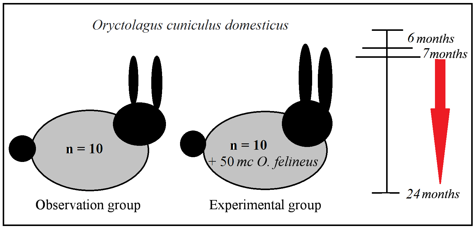
Figure 1. Experimental design. 1-6 months – Intact period (preparing animals for the experiment); 6 months – Infecting animals in a dose of 50 metacercariae (mc) of Opisthorchis felineus (Rivolta, 1884); 7 months – Confirming the infection by O. felineus; 24 months – Removing animals from the experiment.
The number of host animals in each group was 10, based on bioethical principles of the 3Rs: replacement, reduction, and refinement. These principles require a minimum number of animals necessary to ensure reliable data during histological studies, allowing sample data to be considered representative. The total number of animals was determined by the need to identify general patterns of morphological changes under the same conditions for modeling pathology, similar housing conditions, and identical biometric data. The number of animals and groups for the study corresponds to the Mead’s resource equation method: E=N-B-T, where the number of animals (20 in this study) – 1 minus the number of blocks in the study design (0 in this study) – 1 minus the number of treatment groups – 1 yields an error in the degrees of freedom (E) with a numerical value between 10 and 20. The choice of non-rodents was motivated by the study of the specific effects of O. felineus on the host thymus.
The study complied with the 3Rs principle. Replacement: rabbits were chosen because they are not natural carriers of O. felineus, thereby reducing the use of naturally susceptible species. Reduction: the minimum number of animals (n=10 in each group) was determined using a power analysis. Refinement: to minimize suffering, daily health monitoring, environmental enrichment, and painless euthanasia via decapitation were implemented.
The mean weight of each rabbit at the onset of the experiment was 2.8 kg. The rabbits were bred independently and were housed and monitored at the Urgent Veterinary Care Clinic in Kemerovo, Russia, under a cooperative agreement. At the start of the experiment, all 20 animals were examined by a veterinarian and found to be clinically healthy. Each animal was assigned a numbered ear tag applied to the pinna of the outer ear for individual identification.
Animal were kept in individual cubicles, in accordance with their natural circadian rhythms, on a hard floor with hygienic filler (wood chips). The diet included 1 L of briquetted meadow hay (My Animals, Red Rabbit, Tomsk), 150 g of grain (feed wheat) from the Happy Farmer social warehouse, and bottled water Elite Aqua. The animals were prepared for the experiment in advance and received an anthelmintic (Azinox suspension, Agroverzachshita, St. Petersburg, Russia) at a dosage calculated for their weight. Before the experiment, the animals underwent a control stool test for ova and parasites. Animals with feces that did not contain parasite eggs or fragments were selected for the experiment. Domestic rabbits were not previously used as an experimental model of opisthorchiasis. The choice was based on the animal size (not a rodent), its gastrointestinal tract structure (similar to that of humans), its high sensitivity to toxic and infectious agents, its low economic cost, its ease of care and maintenance, and its ethical standards (it is not a pet). A disadvantage of choosing this animal species is the potential for divergence in thymus morphology, as O. felineus is not a natural parasite for rabbits.
Preparation and administration of infectious material
To prepare infectious material for the host animals, I obtained dead fish caught in the Tom River (Tomsk, Russia). The collection of infected fish and their species identification were conducted in collaboration with the Department of Ichthyology and Hydrobiology of Tomsk State University. After transporting the infected fish to Kemerovo, the author manually isolated Siberian liver fluke metacercariae from the fish muscle tissue using a compressor controlled by light microscopy at ×100 magnification. Infection doses of 50 fluke metacercariae were prepared for each definitive host. The infectious material was then administered orally to the animals in the morning on an empty stomach. Completion of the infection was confirmed one month later by positive results of a stool test for O. felineus ova.
Histological material collection and histological methodology
When working with animals, the authors followed the Guide for the Care and Use of Laboratory Animals without inflicting pain, in accordance with the principles of the World Medical Association Declaration on Helsinki (approved by the institutional review board). All applicable international, national, and institutional guidelines for the care and use of animals were followed. All animals were euthanized 18 months after infection (at 24 months of age, at the end of the reproductive period) by immediate painless decapitation. All 20 animals completed the 18-month observation period. No animals died or were excluded from the analysis. No analgesics or anesthesia were administered prior to decapitation, as the procedure is immediate and causes no discomfort when performed correctly by trained personnel. The euthanasia (decapitation) method complies with the American Veterinary Medical Association Guidelines for the Euthanasia of Animals. The study protocol including primary endpoints (number of thymic lobules, specific volumes of thymic cortex and medulla, and number Hassall’s corpuscles) and secondary endpoints (vascular density, lipomatosis of thymic trabeculae) was developed on November 15, 2021, prior to animal allocation to groups.
After confirmation of biological death, autopsy specimens were obtained. Thymus sections (0.5 cm) were immediately fixed in 10% neutral formalin and refixed after 24 hours. Histological examination was performed after a specified time interval. Autopsy specimens were rinsed with tap water and dehydrated in ethanol at increasing concentrations: 40, 50, 60, 70, 80, 90, and 96 degrees, for 60 min each. The samples were then placed in two portions of xylene for 40 min each, after which they were placed in slurry at 37.5 ºС in an incubator for 30 min. The specimens were then compacted in two portions of Histomyx® embedding medium (Biovitrum LLC, Moscow, Russia) for 45 min each in an incubator at 56 ºС. Blocks were prepared and mounted on wooden supports. Then, using a sliding microtome (Kharkiv Medical Equipment Plant, Russia) and disposable microtome blades (Biovitrum LLC, Moscow, Russia), 5-μm-thick organ sections were cut and aligned in a tissue floating bath. The sections were then manually mounted onto glass slides (MiniMed, Russia). After removing Histomyx® from the sections, the latter were stained with Carazzi’s hematoxylin solution and an alcoholic eosin solution. The stains were prepared from powdered reagents manufactured by the State Chemical Committee, SoyuzReaktiv, Russia. Van Gieson staining was employed to identify collagen fibers in connective tissue and assess blood vessels in the thymus. Serial organ sections were prepared, mounting 3-5 sections per slide, and covered with 24×24 mm coverslips (ApexLab, Russia). BioMount (Milan, Italy) was used as the mounting medium.
Research methods
Histological specimens were examined using light microscopy at magnifications of ×40 (0.10), ×100 (0.25), ×400 (0.65), and ×1,000 (1.25) using a Primo Star light microscope (Zeiss, Germany). A descriptive morphological study of the thymus specimens was conducted. Areas with good structural visualization in each specimen were examined, randomly selecting 4-6 fields of view (FOVs) in the cortex, 2-4 FOVs in the medulla depending on the lobule size, or examining the entire specimen due to its small size. Morphometric analysis was performed on specimen images acquired with the built-in camera of the Zeiss Primo Cam microscope using Zen Blue Edition morphometric software (Carl Zeiss, Germany). After scaling, a correction factor was calculated for each magnification to represent the data in actual micrometers. Twenty morphometric measurements were made on specimens from each host animal. The area of organ lobules, as well as their linear dimensions, were measured at a magnification of ×40 (eyepiece magnification ×10, zoom lens ×4).
The organ’s cellular composition was assessed at a magnification of ×1,000 using immersion microscopy, and the cell population density was estimated using the Avtandilov ocular grid. The specific volumes of organ parts and tissue types were calculated using a 100-point ocular grid at a magnification of ×100. The specific volume of blood vessels was calculated at a magnification of ×400 for the thymic cortex and medulla separately. The numbers of lobules and trabeculae in the specimen on serial microsections were calculated at a magnification of ×40. Hassall’s corpuscles (number and structure) were estimated at a magnification of ×400. For each parameter, 20 measurements were taken in different FOVs on specimens obtained from each host animal. Histological preparation, morphometric analysis, and statistical analysis were performed by researchers blinded to group assignment. Specimens were coded and decoded only after all measurements were completed. Statistical processing of the results was performed using Microsoft Office Excel, Statistica software (version 10). Numerical parameter data are presented as the arithmetic mean with standard error of the mean (M±m). Since quantitative morphological characteristics were not normally distributed, they are additionally described by the median (Me) and interquartile range (25th and 75th percentiles), with measures of data dispersion (SD) indicated. Between-group data were assessed using the nonparametric Mann-Whitney U test for samples with a non-normal distribution at a significance level of p≤0.05.
This study was conducted in accordance with ARRIVE 2.0 guidelines (https://arriveguidelines.org) and the Guide for the Care and Use of Laboratory Animals (8th edition, National Research Council).
Results
Histological examination of thymus preparations from animals in the experimental group revealed a number of morphological differences presented in Table 1. Thymic lobules varied in size and shape, with clear morphological boundaries between the cortex and medulla. The boundary of the corticomedullary junction in the thymus of the experimental group animals was indistinct.
Table 1. General morphological characteristics of the thymus in animals with opisthorchiasis
|
## |
Morphological parameter |
Observation group М±m, SD, Ме [25; 75] |
Experimental group М±m, SD, Ме [25; 75] |
Statistical criterion (U; p) * |
|
1 |
Total number of lobules (units)
|
32.55±4.91 4.915 33 [30; 35] |
21.3±5.76 5.768 19.5 [17.5; 25] |
U=33; p=0.000007 |
|
2 |
Specific volume of the cortex (%) |
49±3.74 4.292 49 [47.5; 52] |
23.6±4.1 2.624 24 [22; 25] |
U=0; p=0.000016 |
|
3 |
Specific volume of the medulla (%) |
17.6±2.6 2.062 17 [16; 19.5] |
13.6±1.78 2.981 13.5 [11.5; 15.5] |
U=0; p=0.000183 |
|
4 |
Ratio of the specific volume of the medulla to the cortex (%) |
1:2.78 |
1:1.73 |
- |
|
5 |
Number of connective tissue trabeculae (units) |
13.8±6.14 1.936 14 [12.5; 15] |
9.2±4.78 4.786 8 [5; 12] |
U=109.5; p=0.01493 |
|
6 |
Number of trabeculae with lipomatosis (units) |
22.35±7.4 1.814 22 [21.5; 23] |
7.2±2.98 2.984 7 [5; 10] |
U=19.5; p=0.000001 |
|
7 |
Specific volume of adipose tissue in trabeculae (%) |
1.33±0.57 1.031 1 [0.5; 2] |
0.44±0.1 0.501 0 [0; 1] |
U=0; p=0.00000 |
|
8 |
Specific volume of blood vessels in the thymic cortex (%) |
14.15±4.73 1.496 14 [13; 15] |
8.35±2.56 2.56 8 [6.5; 9.5] |
U=44.5; p=0.000028 |
|
9 |
Specific volume of blood vessels in the thymic medulla (%) |
6.65±2.03 1.725 7 [5.5; 8] |
19.95±7.11 7.112 21 [15; 24] |
U=18; p=0.000001 |
|
10 |
Number of Hassall’s corpuscles in the medulla (units) |
4.05±1.79 2.328 4.5 [1.5; 6] |
7.15±2.15 1.565 7 [6; 8.5] |
U=41.5; p=0.000019 |
|
11 |
Number of Hassall’s corpuscles in the cortex (units) |
- |
1.25±0.14 0.55 1 [0.5; 2] |
- |
Quantitative analysis of lobules in serial organ sections revealed a 1.52-fold decrease in their size in the experimental group vs. the observation group, with statistically significant differences observed between the groups (Table 1, Figure 2). The decrease in organ volume is confirmed by the specific volumes of the cortex and medulla (Table 1). E.g., the specific volume of the cortex was significantly lower in the experimental group than in the observation group. A decrease in the specific volume of the medulla was also observed in the thymus lobules in the experimental group. Moreover, the overall ratio of the specific volume of the medulla to the volume of the cortex is characterized by a threefold shift in favor of the latter, whereas in the observation group, this ratio only slightly exceeds 1.5.
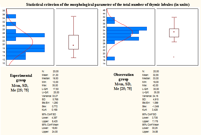
Figure 2. Analysis of the total number of thymus lobules under normal conditions and in opisthorchiasis.
Morphometric analysis of the thymus revealed a decrease in all parameters in the experimental group vs. the observation group (Table 2). Lower values were noted for the total area, cortex, and medulla. Given the smaller area of the thymus lobules in the experimental group, it was appropriate to compare the areas of the lobules in each group with each other. In the experimental group, the area of the medulla was smaller than the area of the cortex. Similar data were obtained in the observation group. The mean total length of the thymus lobule, measured as a linear parameter in one plane, allowed us to spatially obtain its mean value on serial sections of the organ. However, this parameter was also significantly lower than the baseline values by 1.75 times. In particular, the individual lengths of the cortex and medulla in the experimental group were also lower than the baseline values. Taking into account the different thymus sizes in the groups, the ratio of the lengths of the cortex and medulla of the lobules was examined. In both groups, the length of the cortex was greater than the length of the medulla.
Table 2. Morphometric parameters of the thymus in animals with opisthorchiasis
|
## |
Morphometric parameter |
Observation group М±m, SD, Ме [25; 75] |
Experimental group М±m, SD, Ме [25; 75] |
Statistical criterion (U; p) * |
|
1 |
Total lobule area, µm² |
499142.7±195553.3 195553, 447292 [370591; 584232] |
288018.4±142127.5 142127, 247266 [173635; 391001] |
U=71; p = 0.0005 |
|
2 |
Total cortex area, µm² |
406885.9±165350.4 184866, 369125 [299648; 441651] |
226412.1±111035.9 111036, 200881 [139360; 330891] |
U=104; p = 0.009787 |
|
3 |
Total medulla area, µm² |
96652.8±48838.87 48839, 74257 [62096; 142581] |
61606.3±37943.73 37944, 47348 [38857; 68945] |
U=86; p = 0.002139 |
|
4 |
Medulla to cortex area ratio |
1:4.21 |
1:3.67 |
- |
|
5 |
Mean lobular length, µm |
922.81±241.19 241, 896 [777; 1076] |
525.71±180.31 180, 495 [394; 618] |
U=35; p = 0.000009 |
|
6 |
Cortical length (by largest linear dimension), µm |
509.9±120.03 120, 532 [413; 563] |
284.94±132.94 133, 272 [195; 328] |
U=64; p = 0.000247 |
|
7 |
Medullary length (by largest linear dimension), μm |
412.9±179.44 179, 375 [268; 535] |
240.77±108.2 108, 214 [184; 259] |
U=35; p = 0.000009 |
|
8 |
Medulla to cortex length ratio |
1:1.23 |
1:1.18 |
- |
Blood vessels in the thymic cortex in the experimental group were evenly distributed across the entire surface. Their small caliber is noteworthy, as their vascular pattern was primarily characterized by capillary connections, making it difficult to calculate the specific vessel density in the cortex. The number of blood-filled or isolated vascular areas was recorded. Vessels in the subcapsular zone of the cortex, beneath the capsule, and at the origin of the trabeculae had varying blood volumes and were generally dilated and hyperemic. Their identification was straightforward due to the well-developed adventitia. The most dilated blood vessels were observed at the junction of the cortex and medulla – in the area of the corticomedullary junction.
In the medulla, blood vessels were located mainly along the periphery, while they were virtually absent in the central region. It is worth noting that connective tissue in the membranes of the medullary vessels was poorly developed and, in some places, absent. Their proportion in the thymic cortex in experimental group animals was significantly lower by 1.69 times than in the observation group. Conversely, this parameter value in the medulla of the experimental group was significantly higher (threefold) than in the observation group.
In most trabeculae, connective tissue was present only near the thymus capsule, subsequently replaced by adipose tissue. Lipomatous trabeculae (LT) were considered an indicator of age-related organ restructuring (Figure 3). In LT, adipose tissue was arranged in islands or completely replaced them, separating the thymic lobules.
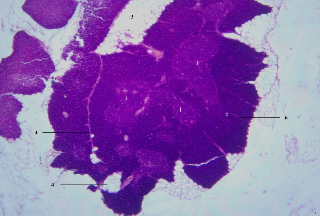
Figure 3. The thymus of a rabbit in the chronic phase of opisthorchiasis after 1.5 years. Staining: hematoxylin and eosin. Light microscopy, magnification: ×40. 1: Thymic medulla; 2: Thymic cortex; 3: White adipose tissue; 4: Trabeculae containing islands of white adipose tissue and individual adipocytes (trabeculae with lipomatosis); 5: Thymic lobule; 6: Trabeculae consisting of connective tissue. Scale bar = 100 µm.
Other trabeculae, in which adipose tissue was not visualized in microsections, contained only connective tissue and were considered structures without age-related restructuring (SWAR).
The number of SWAR in the experimental group was statistically significantly lower than baseline values (by 1.5 times). Moreover, the number of lipomatous trabeculae was 3.1 times lower in the experimental group compared to baseline values.
The specific volume of adipose tissue in thymic trabeculae in the observation group was approximately threefold of that in the experimental group.
When comparing parameters within the same group (SWAR/LT), a predominance of LT was observed in the observation group (1:1.61). However, when comparing the same parameters in the experimental group, the reverse pattern was observed: 1.27:1. Therefore, age-related restructuring of thymic trabeculae was more pronounced in the observation group than in the experimental group.
Hassall’s corpuscles were detected in the thymic cortex and medulla in the experimental group (Figures 4 and 5). Typical (mature) Hassall’s corpuscles with a formed keratin core were present in the thymic cortex, which is quite rare. All types were present in the medulla, including progressive, degenerative, and mature types. Mast cells were detected in the outer layer of concentric corpuscles in the thymic medulla. The number of Hassall’s corpuscles in the thymic medulla of the experimental group animals was significantly higher (1.76 times) than in the observation group. No Hassall’s corpuscles were detected in the thymic cortex of the observation group.
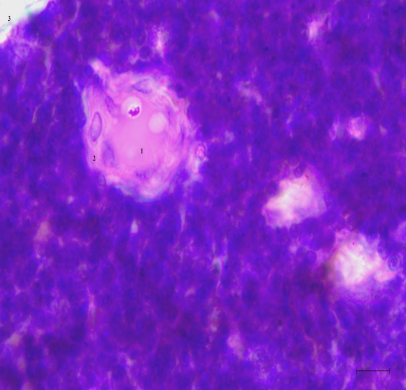
Figure 4. Hassall’s corpuscles in the thymic cortex of a rabbit in the chronic phase of opisthorchiasis after 1.5 years. Staining: hematoxylin and eosin. Light immersion microscopy, magnification: ×1,000. 1: Degenerative reticular epithelial cell in the center of a Hassall’s corpuscle in the thymic cortex; 2: Flattened reticular epithelial cells; 3: Thymus capsule.
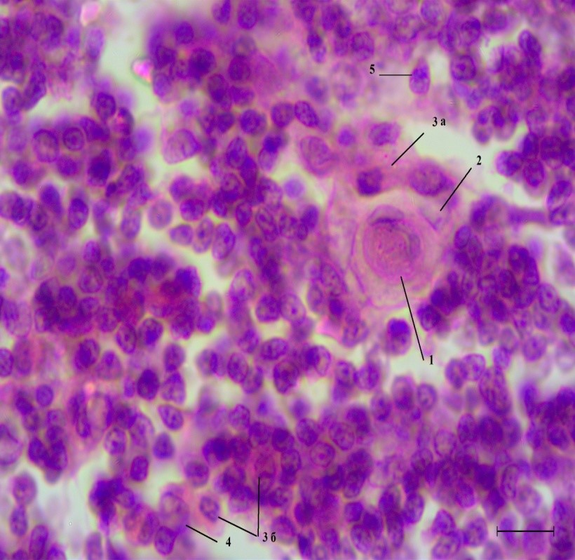
Figure 5. Hassall’s corpuscles in the thymic medulla of a rabbit in the chronic phase of opisthorchiasis after 1.5 years. Staining: hematoxylin and eosin. Light immersion microscopy, magnification: ×1,000. 1: Degenerative reticular epithelial cell in the center of a Hassall’s corpuscle; 2: Flattened reticular epithelial cells; 3a: Mast cell within a Hassall’s corpuscle; 3b: Mast cell in the medulla; 4: Reticular epithelial cell of the medulla; 5: Macrophage. Scale bar = 10 µm.
The cellular composition in the thymic cortex revealed by the light immersion microscopy, was characterized by a dense population of cellular elements consisting of reticular epithelial cells of the organ’s stroma, maturing forms of lymphocytes, mast cells, macrophages, and monocytes (Figure 6). Mature lymphocytes located near the corticomedullary border and lymphoblasts in the subcapsular region were visible in the cortex, while other forms of maturing lymphocytes could not be identified. Mast cells and monocytes were localized in the subcapsular zone, immediately behind the connective tissue capsule or along the origin of trabeculae descending from the capsule between the organ lobules. Mast cells were characterized by their substantial size, oval or round nuclei, and characteristic granular cytoplasm. Macrophages were found throughout the cortex. In the corticomedullary zone, macrophages were characterized by the presence of phagocytic bodies of varying shapes in their cytoplasm. In both groups, reticular stromal epithelial cells and mature lymphocytes were present in the medulla.
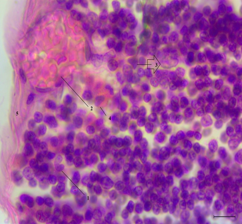
Figure 6. Cells in the subcapsular zone of the thymic cortex of a rabbit in the chronic phase of opisthorchiasis after 1.5 years. Staining: hematoxylin and eosin. Light immersion microscopy, magnification: ×1,000. 1: Monocyte; 2: Blood vessel; 3: Reticular epithelial cell; 4: Mast cell; 5: Thymus capsule. Scale bar = 10 µm.
Discussion
An analysis of morphological and morphometric changes in the thymus of ontogenetically post-reproductive animals via an experimental model of opisthorchiasis revealed significant differences. The values of many studied morphological parameters of the thymus were significantly lower during opisthorchiasis infection vs. the baseline level, which should be considered a sign of age-related organ transformation occurring earlier. According to current publications, age-related thymic involution happens due to the degeneration of the thymic epithelium [Fujimori S. et al., 2024]. Age-related thymic involution leads to a gradual thymic cellularity decline, a decrease in the number of reticular epithelial cells of the cortex and medulla, along with an increase in the number of fibroblasts and, consequently, to organ atrophy and its replacement with connective tissue [Liang Z. et al., 2022]. The thymus shrinks in size after the first year of life, annually accounting for 3% of the total organ mass, while the boundary between the cortex and medulla becomes indistinct [Liang Z., et al., 2022]. In a rabbit with opisthorchiasis, a decrease in the number of lobules, in the specific volume of the cortex and medulla, and in the number of trabeculae was observed. These parameters collectively account for a decrease in the total volume of the thymus, which can be considered atrophy, compared with the baseline. Indistinct boundary between the cortex and medulla was also noted, further indicating organ atrophy in opisthorchiasis.
A reliable indicator of the functional state of the thymus is the ratio of its cortex to medulla [Kreins A.Y. et al., 2020]. Parasitic diseases trigger organ atrophy, but the cortex is primarily affected, while the medulla is characterized by hypertrophy [Kozlov V.A., 2019]. However, with opisthorchiasis, a slight tendency toward organ rejuvenation was also observed, as the cortex-to-medulla ratio is virtually identical, while the reverse trend was noted in the observation group.
However, a closer examination of the morphological features revealed that signs of lipomatosis in the thymus of the experimental group were less pronounced. As the thymus ages, stromal sclerosis and lipomatosis of the interlobular trabeculae develop [Molchanova A.A. et al., 2017]. The biological age of the animals in this study corresponded to the completion of the reproductive period of ontogenesis, and the housing conditions in both groups were identical, indicating that morphological changes in the thymus occurred as a result of parasitic infection. With O. felineus infection, the thymic lobes and the ratio of the medulla to cortex were found to be smaller, compared with the baseline, implying a reduction in the cortex volume. In the experimental group, I observed a reduction in the number of thymic lobules and trabeculae (interlobular connective tissue) observed in serial organ sections, and in the specific volumes of the medulla and cortex, as well as in the vascular component. This reduction was due to an overall decrease in organ volume. These characteristics suggest that age-related thymic involution occurs to a greater extent in cases of simultaneous parasitic disease and natural aging.
Yet, other morphometric parameters contradict this conclusion, because animals in the experimental group did not exhibit the same volume of parenchyma replacement with adipose tissue. The specific volume of adipose tissue was insignificant, and there were few LT. The number of LT in opisthorchiasis was insignificant, and the specific volume of adipose tissue was lower than baseline, which can be interpreted as a deceleration of organ aging. Such morphological changes are likely due to factors secreted by the Siberian liver fluke. During opisthorchiasis, parthenitae secrete extracellular vesicles containing ferritin, tetraspanin CD63, helminth defense molecule-1, globin-3, saposin B domain-containing protein, 60S ribosomal protein, glutathione S-transferase class-mu 28 kDa isozyme (GST28), tubulin, and thioredoxin peroxidase, known as promoters of cholangiocyte neoplasia [Pakharukova M.Yu., 2023].
With age, the thymic stromal microenvironment is disrupted by the loss of some corticomedullary junctions and a decrease in the number of reticular epithelial cells in the cortex and medulla, fibroblast proliferation, and an increase in perivascular space [Liang Z. et al., 2022]. Stimulation of collagenogenesis by fibroblasts in thymic trabeculae during opisthorchiasis may likely be caused by tissue hypoxia due to a decrease in the number of blood vessels, which requires further study.
Different specific volumes of blood vessels in the thymic cortex and medulla in the experimental group characterize the differentiated impact of parasitic infection on lobular components. A significant number of blood-filled vessels indicates increased blood supply to a specific area of the organ, viz., the medulla. The recirculating pool of T lymphocytes in the thymic medulla may play a role in the humoral feedback loop between peripheral and central organs of immunogenesis. The parasite likely produces vasoactive factors that exert a relaxant effect on smooth muscle cells in the vessel walls, which requires further study. Increased concentrations of these factors in the chronic phase results in congestion in the organ, manifested as vascular hyperemia, visualized in sections. In the cortex, such factors may be degraded by reticular epithelial cells or may lack target cells. The decrease in vascularization may be associated with enhanced self-protection of the blood-thymus barrier, aimed at reducing the number of antigens delivered to naïve lymphocytes.
Morphological changes in the thymus during parasitic diseases often exhibit signs of acute thymic involution, since parasitism is a long-term process requiring constant adaptation to changing conditions. The parasite exerts local mechanical, toxic, and other effects on the bile duct walls. However, with mechanical injuries characterized by lengthy wound healing process, dynamic changes in the cortex and medulla of the thymic lobules occur later in life [Khalikov A.A. et al., 2021].
Parasitic transformation (involution) of the thymus in opisthorchiasis has an atypical morphological picture, combining the features of both age-related and acute thymic involution that manifest as a consequence of pathological changes and regenerative processes. However, a distinctive feature of parasitic thymic involution is the presence of subtle signs, such as a certain loss of corticomedullary differentiation and the absence of cortical depletion, despite the presence of isolated concentric corpuscles. Siberian liver fluke has been shown to secrete proliferative factors [Mordvinov V.A. et al., 2020], which can reach the thymus via lymphogenous and/or hematogenous routes. Consequently, in opisthorchiasis, acute thymic involution is initiated but immediately suppressed, accompanied by signs of organ regeneration. As a result, immunosuppression in parasitic diseases initially develops with the release of large quantities of antigens by the parasite, followed by post-involutional regeneration under the influence of factors secreted by the parasite.
In other helminthiases, research data are characterized by certain common features of morphological organ transformation. For example, in the case of infection with Diphyllobothrium dendriticum, sclerotic lesions of the organ stroma, inversion of lobular layers into each other, an increase in the size and number of Hassall’s corpuscles, as well as a 1.5-fold increase in the medullary content in the lobules, implying organ involution, are present in the thymus of golden hamsters by Day after the infection [Pronina S.V. et al., 2009]. Moreover, in mice with leishmaniasis, thymic hypertrophy was observed, including cortical thickening on Day 3 and loss of corticomedullary differentiation on Day 7 after the infection [Arrais-Lima C. et al., 2021]. Toxoplasma gondii infection results in thymic atrophy, the degree of which depends on the pathogen’s virulence [Kozlov V.A., 2019]. Consequently, morphological changes in the thymus vary depending on the type of infection and the definitive host. Gastrointestinal lumen parasites tend to cause involutional modifications, while intracellular parasites tend to cause hyperplastic changes. Furthermore, intestinal and intrahepatic parasites appear to have different effects on the thymus, the mechanisms of which require further comparative analysis.
Parasites exert a powerful immunosuppressive effect on the host to remain undetected by the immune system, but these mechanisms appear to lose their effectiveness as the duration of parasitic infection increases and are replaced by other adaptation processes. E.g., acute infection with O. felineus is accompanied by the induction of B lymphocytes and the suppression of T lymphocyte populations (CD4+, CD8+) [Litvinova L.S. et al., 2006]. T lymphocyte deficiency was observed as a result of their massive apoptosis in the spleen caused by proteasome dysfunction, but not in the thymus [Shinebaatar E. et al., 2025]. Immunosuppression in opisthorchiasis, as in other helminthiases, is biologically significant for the survival of the parasite in the host organism, since it depends on many factors, including genetic tolerance, immune system reactivity, infective dose, and the presence of parasitic superinfection. I did not observe a loss of corticomedullary differentiation or a decrease in the cellularity of the cortex in the thymus during opisthorchiasis, which indicates active organ function. However, further studies of the immunological profile of thymic lymphocytes are necessary to interpret organ function. For instance, during parasitic infection, the immune system counteracts the proliferation of parasitic forms. However, for O. felineus, the number of parthenitae depends on the number of metacercariae consumed, provided they survive in the definitive host. Furthermore, thymic factors may influence increased egg production in Siberian liver fluke, which requires further study.
Hassall’s corpuscles were significantly more common in the thymus of the experimental group animals, reflecting the stressful impact of fluke infection on the organ. The formation of the corpuscles is due to glucocorticoids during acute thymic involution. Other authors believe that thymus damage occurs systemically due to the influence of other damaged organs and the associated release of glucocorticoids and proinflammatory molecules [Luo M. et al., 2020]. Sex steroids induce early age-related thymic involution. Testosterone supplementation of castrated animals yields early thymus atrophy and lymphocyte apoptosis, suggesting that hormonal changes directly drive organ remodeling [Li D. et al., 2024]. Morphological indices of age-related thymic changes in males of both groups differed statistically significantly, possibly due to differences in gonadal endocrine function.
The presence of concentric corpuscles (Hassall’s corpuscles) in the thymic cortex and at the corticomedullary junction during O. felineus infection can be considered a sign of acute thymic involution. Their presence in the cortex and an increase in their numbers in the medulla indicate the chronic phase of parasitic infection, which is a noteworthy stress factor for the body. Their appearance in the thymus is usually influenced by glucocorticoids secreted by the adrenal glands. The mechanism of formation of these corpuscles during opisthorchiasis is likely also related to the release of cortisol. Glucocorticoid release by the adrenal glands is not a pathogenic factor in opisthorchiasis, but their production during adaptation to infection is important as potent immunosuppressants, inhibiting inflammation via anti-inflammatory cytokines (e.g., IL-10). During opisthorchiasis, thymocytes produce key proinflammatory cytokines (IL-1β, IL-6, TNF-α, and IL-8), which stimulate the development of chronic inflammation, including cholangitis.
According to the published sources, thymus functions are controlled by autocrine/paracrine regulatory mechanisms mediated by hormones and neuropeptides, which can be altered under conditions of infectious stress and chronic inflammation, as observed during Trypanosoma cruzi infection. [González F.B. et al., 2024]. The formation of a larger number of concentric corpuscles is likely important for adaptive mechanisms in the host–parasite system. During cellular interactions, reticular epithelial cells produce numerous hormones (factors) known for their antitumor activity, which may explain the large number of concentric corpuscles in all parts of the lobules. It is possible that an increase in the number of concentric corpuscles is necessary to maintain effective antiparasitic immune response.
With age, the number of Hassall’s corpuscles decreases, which, according to the authors, occurs due to a reduced influx of bone marrow precursors and a reduction in the number of reticular epithelial cells [Erofeeva L.M., 2017]. Physiological and reparative types of thymus regeneration have their own characteristics, making it impossible to slow or even interrupt age-related organ restructuring [Postovalova E.A. et al., 2019]. Consequently, during O. felineus infection, organ aging is decelerated by its augmented stimulation, which increases blood flow within the organ and stimulates T cell production through feedback mechanisms from peripheral organs under conditions of functional stress.
At the same time, compensatory stimulation of lymphocyte production likely also occurs in another central organ. E.g., the red bone marrow is closely interconnected with the thymus and is also susceptible to disease triggers, despite the presence of a blood-tissue barrier. Infection of rabbits with O. felineus is accompanied by a disruption in the formation of maturing cell forms, manifested by a delay in the appearance of cytoplasmic structures and a change in the shape of the nucleus, while stromal components are preserved [Sidelnikova A.A., 2018].
Intracellular parasitic infection with T. cruzi in Chagas disease leads to temporary suppression of red bone marrow immunity, a decrease in its cellularity, malfunctioning development of thymocytes, and thymus atrophy due to a decrease in the number of common lymphoid progenitors (CLPs) and lymphoid-primed multipotent progenitors (LMPPs) [Marins-Dos-Santos A. et al., 2022]. This means that the parasite’s effects are not limited to localized organs but also affect other systems, particularly those with an active blood supply. One such organ is the kidney, where species-specific reactions (periductal fibrosis) are observed during infection with O. felineus and Clonorchis sinensis [Zaparina O.G. et al., 2024]. In contrast, no fibrosis development in the thymus was observed during the observation period in the study; perhaps a longer observation period is necessary for its development.
A comparative assessment of the morphological features of thymic involutions (transformations) is presented in Table 3. Data on acute thymic involution were taken from the literature and used for the comparative assessment. The table shows that the animals of the observation group exhibited more pronounced signs of age-related involution, while parasitic transformation (involution) of the organ combined various features expressed to varying degrees, emphasizing its uniqueness.
Table 3. Comparative matrix of thymus transformation (involution) types
|
Morphological parameter |
Observation group: age-related thymic involution |
Experimental group: parasitic transformation (involution) |
Acute thymic involution: comparison based on literature data |
|
Loss of the cortex-medulla boundary |
+ Very weak |
+ Weak |
+ Very pronounced |
|
Presence of Hassall’s bodies in the cortex |
– |
+ Obvious |
+ Very pronounced |
|
Presence of Hassall’s bodies in the medulla |
+ Weak |
+ Obvious |
+ Very pronounced |
|
Total number of lobules (decrease in number) |
+ Very pronounced |
+ Pronounced |
– |
|
Total area of lobules (decrease in size) |
+ Very pronounced |
+ Pronounced |
– |
|
Specific volume of adipose tissue in the organ |
+ Very pronounced |
+ Very weak |
– |
|
Number of trabeculae with lipomatosis |
+ Pronounced |
+ Weak |
– |
|
Specific volume of blood vessels |
Weak |
+ Very pronounced |
+ Pronounced |
Hence, only a few morphological criteria were identified, while the role of the thymus in the development of the antiparasitic response requires further study through dynamic observation, detailed endocrine analysis, morphological and cytological parameters, as well as the potential and limitations of age-related and acute thymic involutions.
Conclusion
A study of morphological changes in the thymus following parasitic infection by O. felineus revealed uneven intraorgan structural changes, more characteristic of the organ parenchyma than of stromal components. Morphological signs of acute thymic involution following parasitic infection by O. felineus are moderately pronounced. The thymus in animals infected with O. felineus is characterized by conflicting signs of both age-related and acute involution. The combination of these signs and signs of organ regeneration suggests a specific type of organ change – parasitic transformation (involution).
Limitations of the study
Potential limitations of the study include the detailed analysis of organ changes during the maximum observation period of infection, due to the short lifespan of the animals, as well as the impossibility of continuous observation of morphological changes in the organ over time. Only point-by-point time-course analysis of histological material during infection was performed, which is an acceptable comparison with similar morphological changes in humans.
Acknowledgments
The author is thankful to the faculty of Kemerovo State Medical University (Russia) S.F. Zinchuk, PhD, and A.I. Vavilov, MD, for training in parasitological research methods and providing material for the study (fish caught in the Tom River, Tomsk). Sincere gratitude is expressed to E.A. Interesova, DSc, Laboratory of Ichthyology Research, Novosibirsk Branch of the Russian Federal Research Institute of Fisheries and Oceanography, for organizing the experiment. The input by L.V. Nacheva, DSc, is very much appreciated: she provided the receipt of the research material and participated in the study of opisthorchiasis.
Conflict of interest
None declared by the author
AI statement
I did not use any artificial intelligence tools or technologies to prepare this manuscript.
- Erofeeva LM. The age changes of the tissue structure and the cellular composition of the human thymus. Morphological Newsletter 2017; 25(2): 21-26. Russian. https://doi.org/10.20340/mv-mn.17(25).02.03.
- Zabrodin VA. Anatomical nomenclature and terminology of the thymus. Vestnik of the Smolensk State Medical Academy 2003; (4): 15-17. Russian. https://sgma.info/en/archive/2000-2009/2003/2003-4.html.
- Ivanovskaya TE, Zairatyants OV, Leonova LV, Voloshchuk IN. Pathology of the Thymus in Children. St. Petersburg: Sotis. 1996: 270-272. Russian. https://search.rsl.ru/ru/record/01001745681.
- Kozlov VA. T cell thymic selection and peripheral homeostatic proliferation in infectious diseases. Russian Journal of Infection and Immunity. 2019; 9(5-6): 629-638. Russian. https://doi.org/10.15789/2220-7619-2019-5-6-629-638.
- Litvinova LS, Kolobovnikova YuV, Knutareva EN, Grigorieva ES, Suvorova EV, Novitsky VV, et al. Cytotoxic potential of eosinophilic granulocytes in patients with eosinophilia syndrome. Bulletin of Siberian Medicine 2006; 5(3): 26-30. https://elibrary.ru/hubdrl.
- Mordvinov VA, Ershov NI, Zaparina OG, Pakharukova MYu. Genomics and proteomics of the liver fluke Opisthorchis felineus. Vavilov Journal of Genetics and Breeding 2020; 24(4): 383-390. Russian. https://doi.org/10.18699/VJ20.44-o.
- Molchanova AA, Grinberg VB, Kushakov KT. A thymus role in pathology of adults (Review). Vestnik Almatinskogo Gosudarstvennogo Instituta Usovershenstvovaniia Vrachei 2017; (3): 17-22. Russian. https://elibrary.ru/ylpwhv.
- Pronina SV, Fomina AS. Morphofunctional changes in the hamster’s thymus testify about strong immunological reaction to casual host which caused by high level of antigen reaction of Diphyllobothrium dendriticum in a stage of active growth. Biulleten Vostochno-Sibirskogo Nauchnogo Tsentra Sibirskogo Otdeleniia Rossiiskoi Akademii Meditsinskikh Nauk 2009; (2): 125-128. Russian. https://elibrary.ru/mqoxwr.
- Postovalova EA, Makarova OV, Kosyreva AM, Mikhailova LP. Morphology of the thymus and the specific features of its cellular composition in experimental acute and chronic ulcerative colitis. Archive of Pathology 2019; 81(5): 53-63. Russian. https://doi.org/10.17116/patol20198105153.
- Sidelnikova AA. Morphological changes of bone marrow in the experimental opisthorchiasis. Morphological Newsletter 2018; 26(1):55-57. Russian. https://doi.org/10.20340/mv-mn.18(26).01.55-57.
- Khalikov AA, Kildyushov EM, Kuznetsov KO, Komlev DS, Rahmatullina GR. Possibility of determining injury duration based on changes in histomorphometric characteristics of the thymus. Russian Journal of Forensic Medicine 2021; 7(2): 96-100. Russian. https://doi.org/10.17816/fm401.
- Arrais-Lima C, Lima LR, Faria MGBF, Degrossoli A, Arrais-Silva WW, Souto PCS. Morphological alterations of Thymus during the early murine leishmaniasis. Trop Biomed 2021; 38(3): 338-342. https://doi.org/10.47665/tb.38.3.075.
- Cowan JE, Takahama Y, Bhandoola A, Ohigashi I. Postnatal involution and counter-involution of the thymus. Front Immunol 2020. 11: 897. https://doi.org/10.3389/fimmu.2020.00897.
- Fujimori S, Ohigashi I. The role of thymic epithelium in thymus development and age-related thymic involution. J Med Invest 2024; 71(1.2): 29-39. https://doi.org/10.2152/jmi.71.29.
- González FB, Savino W, Pérez AR. Brain-Thymus Connections in Chagas Disease. Neuroimmunomodulation 2024; 31(1): 78-88. https://doi.org/10.1159/000538220.
- Karbysheva N, Nikonorova M, Matros O, Kiushkina I, Nemilostiva E, Choroshilova I, et al. Clinical polymorphism in patients with Opisthorchis felineus infection in the Western Siberia. IDCases 2021; 24: e01064. https://doi.org/10.1016/j.idcr.2021.e01064.
- Kreins AY, Graham Davies E. Replacing defective thymus function. Curr Opin Allergy Clin Immunol 2020; 20(6): 541-548. https://doi.org/10.1097/aci.0000000000000695.
- Liang Z, Dong X, Zhang Z, Zhang Q, Zhao Y. Age-related thymic involution: Mechanisms and functional impact. Aging Cell 2022; 21(8): e13671. https://doi.org/10.1111/acel.13671.
- Li D, Yao H, Ren Y, Shang J, Han X, Cao X, Song T, Zeng X. Testosterone regulates thymic remodeling by altering metabolic reprogramming in male rats. Gen Comp Endocrinol 2024; 348: 114448. https://doi.org/10.1016/j.ygcen.2024.114448.
- Luo M, Xu L, Qian Z, Sun X. Infection-Associated Thymic Atrophy. Front Immunol 2021; 12: 652538. https://doi.org/10.3389/fimmu.2021.652538.
- Marins-Dos-Santos A, Ayres-Silva JP, Antunes D, Moreira CJC, Pelajo-Machado M, Alfaro D, et al. Oral Trypanosoma cruzi Acute Infection in Mice Targets Primary Lymphoid Organs and Triggers Extramedullary Hematopoiesis. Front Cell Infect Microbiol 2022; 12: 800395. https://doi.org/10.3389/fcimb.2022.800395.
- Pakharukova MY, Savina E, Ponomarev DV, Gubanova NV, Zaparina O, Zakirova EG, et al. Proteomic characterization of Opisthorchis felineus exosome-like vesicles and their uptake by human cholangiocytes. J Proteomics 2023; 283-284: 104927. https://doi.org/10.1016/j.jprot.2023.104927.
- Shinebaatar E, Morimoto J, Koga R, Nguyen TN, Sasaki Y, Yonemura S, et al. Proteasome dysfunction in T cells causes immunodeficiency via cell cycle disruption and apoptosis. Int Immunol 2025; 37(8): 493-505. https://doi.org/10.1093/intimm/dxaf021.
- Zaparina OG, Kapushchak YK, Lishai EA, Hong SJ, Sripa B, Pakharukova MY. Species-specific renal and liver responses during infection with food-borne trematodes Opisthorchis felineus, Opisthorchis viverrini, or Clonorchis sinensis. PLoS One 2024; 19(12): e0311481. https://doi.org/10.1371/journal.pone.0311481.
- Zhang WL, Zeng YH, Lai YS. Spatial-temporal risk of Opisthorchis felineus infection in Western Siberia and the Ural Region of Russian Federation: a joint Bayesian modelling study based on survey and surveillance data. Infect Dis Poverty 2025; 14(1): 95. https://doi.org/10.1186/s40249-025-01363-z.
Received 2 June 2025, Revised 19 May 2026, Accepted 5 June 2026
© 2025, Russian Open Medical Journal
Correspondence to Alevtina A. Sidelnikova. Phone: +79236130701. E-mail: alieva-alevtina@mail.ru.